Product Detail
Product NameUbiquitin D Rabbit mAb
Clone No.JJ084-09
Host SpeciesRecombinant Rabbit
Clonality Monoclonal
PurificationProA affinity purified
ApplicationsWB, ICC, IHC
Species ReactivityHu, Ms
Immunogen Descrecombinant protein
ConjugateUnconjugated
Other NamesDiubiquitin antibody FAT10 antibody GABBR1 antibody UBD 3 antibody Ubd antibody UBD_HUMAN antibody Ubiquitin D antibody Ubiquitin like protein FAT10 antibody Ubiquitin-like protein FAT10 antibody
Accession NoSwiss-Prot#:O15205
Uniprot
O15205
Gene ID
10537;
Calculated MW18 kDa
Formulation1*TBS (pH7.4), 1%BSA, 40%Glycerol. Preservative: 0.05% Sodium Azide.
StorageStore at -20˚C
Application Details
WB: 1:1,000-1:2,000
IHC: 1:50-1:200
ICC: 1:100-1:500
Western blot analysis of Ubiquitin D on Hela cells lysates using anti-Ubiquitin D antibody at 1/1,000 dilution.
Immunohistochemical analysis of paraffin-embedded mouse kidney tissue using anti-Ubiquitin D antibody. Counter stained with hematoxylin.
Immunohistochemical analysis of paraffin-embedded human kidney tissue using anti-Ubiquitin D antibody. Counter stained with hematoxylin.
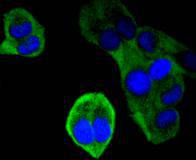
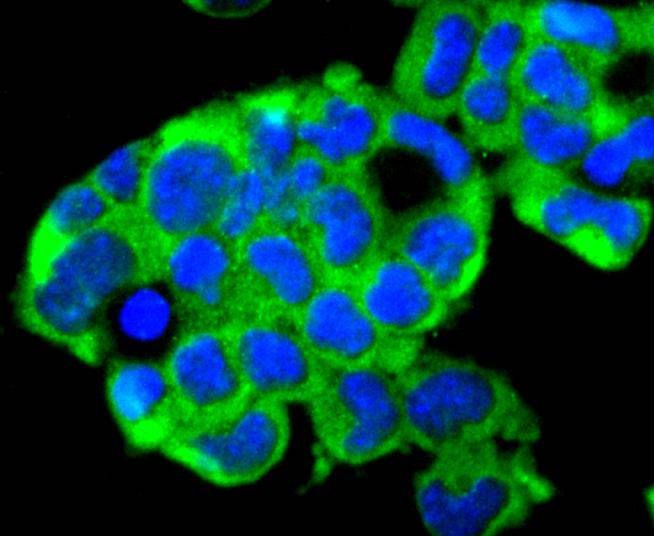
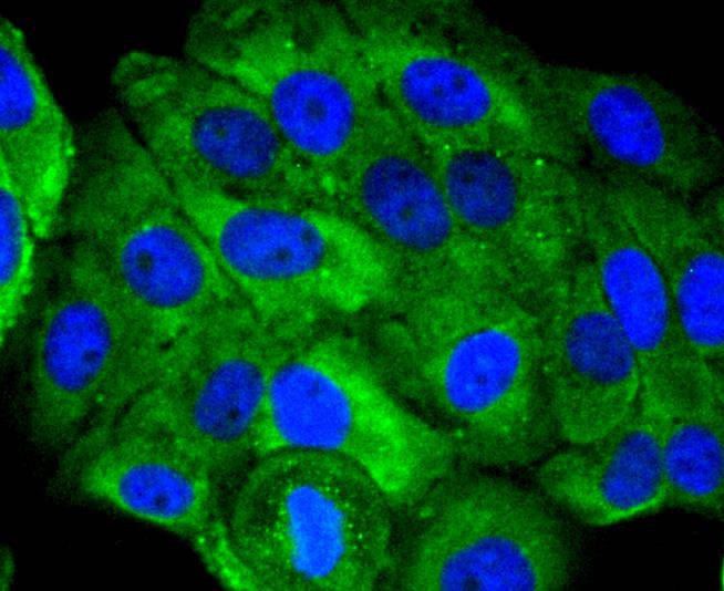
ICC staining Ubiquitin D in Hela cells (green). The nuclear counter stain is DAPI (blue). Cells were fixed in paraformaldehyde, permeabilised with 0.25% Triton X100/PBS.
ICC staining Ubiquitin D in F9 cells (green). The nuclear counter stain is DAPI (blue). Cells were fixed in paraformaldehyde, permeabilised with 0.25% Triton X100/PBS.
ICC staining Ubiquitin D in HepG2 cells (green). The nuclear counter stain is DAPI (blue). Cells were fixed in paraformaldehyde, permeabilised with 0.25% Triton X100/PBS.
FAT10, also designated Ubiquitin D or Diubiquitin, is a 165 amino acid protein encoded in the major histocompatibility complex (MHC) that consists of two domains which share significant homology with ubiquitin. Each domain contains two cysteines, along with a free C-terminal diglycine motif required for FAT10 conjugate formation. FAT10 is inducible by interferon-g and tumor necrosis factor a (TNF?). The FAT10 protein interacts with MAD2, a component of the spindle checkpoint, and plays a role in antigen presentation, cytokine response, apoptosis and mitosis. It may also regulate cell growth during dendritic cell or B cell activation and development. FAT10 mRNA is expressed mainly in some dendritic cells and lymphoblastoid lines and in other specific cells subsequent to interferon-g induction. The human FAT10 gene, designated UBD, maps to chromosome 6p21.3 and is overexpressed in the tumors of various epithelial cancers.
If you have published an article using product 49304, please notify us so that we can cite your literature.


 Yes
Yes


