Welcome to Sabbiotech!     
Email:info@signalwayantibody.com
Location: Home > Recombinant Rabbit Monoclonal Antibodies > PTP1B Rabbit mAb

PTP1B Rabbit mAb#49305

Availability: Yes
Total Amount:$
Product Detail

Product NamePTP1B Rabbit mAb

Clone No.JJ0935

Host SpeciesRecombinant Rabbit

Clonality Monoclonal

PurificationProA affinity purified

ApplicationsWB, ICC/IF, IHC, FC

Species ReactivityHu, zebrafish

Immunogen Descrecombinant protein

ConjugateUnconjugated

Other NamesPTP1B antibody
Non receptor tyrosine phosphatase 1 antibody
Protein phosphotyrosylphosphatase 1B antibody
Protein tyrosine phosphatase 1B antibody
Protein tyrosine phosphatase non receptor type 1 antibody
Protein tyrosine phosphatase placental antibody
Protein-tyrosine phosphatase 1B antibody
PTN1_HUMAN antibody
PTP 1B antibody
PTP-1B antibody
PTPN 1 antibody
PTPN1 antibody
Tyrosine protein phosphatase non receptor type 1 antibody
Tyrosine-protein phosphatase non-receptor type 1 antibody

Accession NoSwiss-Prot#:P18031

Uniprot P18031

Gene ID 5770;

Calculated MW50 kDa

Formulation1*TBS (pH7.4), 1%BSA, 40%Glycerol. Preservative: 0.05% Sodium Azide.

StorageStore at -20˚C

Application Details
WB: 1:1,000-1:2,000
IHC: 1:50-1:200
ICC: 1:100-1:500

FC: 1:50-1:100
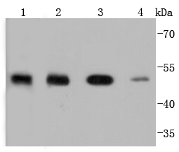
Western blot analysis of PTP1B on different lysates using anti-PTP1B antibody at 1/1,000 dilution. Positive control:
Lane 1: zebrafish
Lane 2: MCF-7
Lane 3: HepG2
Lane 4: A431
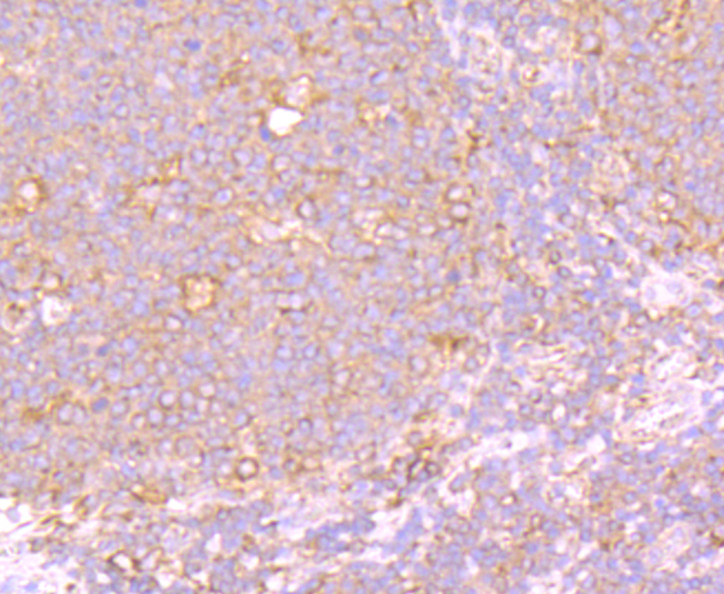
Immunohistochemical analysis of paraffin-embedded human tonsil tissue using anti-PTP1B antibody. Counter stained with hematoxylin.
ICC staining PTP1B in Hela cells (green). The nuclear counter stain is DAPI (blue). Cells were fixed in paraformaldehyde, permeabilised with 0.25% Triton X100/PBS.
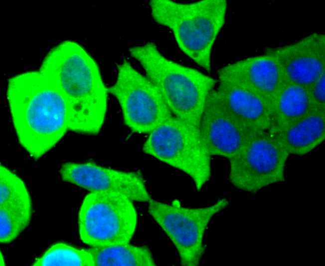
ICC staining PTP1B in MCF-7 cells (green). The nuclear counter stain is DAPI (blue). Cells were fixed in paraformaldehyde, permeabilised with 0.25% Triton X100/PBS.
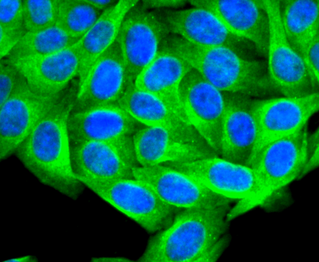
ICC staining PTP1B in HepG2 cells (green). The nuclear counter stain is DAPI (blue). Cells were fixed in paraformaldehyde, permeabilised with 0.25% Triton X100/PBS.
Flow cytometric analysis of Raji cells with PTP1B antibody at 1/50 dilution (red) compared with an unlabelled control (cells without incubation with primary antibody; black). Alexa Fluor 488-conjugated goat anti rabbit IgG was used as the secondary antibody
The phosphorylation of proteins at tyrosine residues has long been recognized as an important regulatory component of signal transduction. This is a reversible process, involving both enzymes that phosphorylate proteins on tyrosine residues as well as a rapidly expanding family of protein tyrosine phosphatases. These latter enzymes bear little resemblance to either the protein serine and protein threonine phosphatases or to the acid and alkaline phosphatases. In most tissues, the major PTPase is a vanadate- and molybdate-sensitive protein. On the basis of sequence analysis, PTP1B (PTPase 1B) expressed in human placenta exhibits similarities both with the common leukocyte antigen (CD45) and with LAR, a homolog of the neural adhesion molecule (NCAM). PTP1B is synthesized as a 435 amino acid precursor protein which is cleaved to generate the active 321 amino acid enzyme.

If you have published an article using product 49305, please notify us so that we can cite your literature.

NOTE

Application

  • WBWestern Blotting
  • IHCImmunohistochemistry
  • IFImmunofluorescence
  • ICCImmunocytochemistry
  • FCFlow Cytometry
  • IPImmunoprecipitation
  • EELISA
  • DBDot Blotting
  • ChIPChromatin Immunoprecipitation
  • GICAGold Immunochromatography Assay
  • NCNegative Control

Species Reactivity

  • HuHuman
  • MsMouse
  • RtRat
  • DmDrosophila melanogaster
  • CCaenorhabditis elegans
  • MkMonkey
  • RbRabbit
  • BBovine
  • DDog
  • PPig
  • HmHamster
  • ChHmChinese Hamster
  • ChkChicken
  • ShpSheep
;