Product Detail
Product NameFas(CD95) Rabbit mAb
Clone No.SR4535
Host SpeciesRecombinant Rabbit
Clonality Monoclonal
PurificationProA affinity purified
ApplicationsWB, ICC/IF, IHC, FC
Species ReactivityHu
Immunogen Descrecombinant protein
ConjugateUnconjugated
Other NamesALPS 1A antibody ALPS1A antibody APO 1 antibody Apo 1 antigen antibody APO 1 cell surface antigen antibody Apo-1 antigen antibody APO1 antibody Apo1 antigen antibody APO1 cell surface antigen antibody Apoptosis antigen 1 antibody Apoptosis mediating surface antigen FAS antibody Apoptosis-mediating surface antigen FAS antibody APT 1 antibody APT1 antibody CD 95 antibody CD 95 antigen antibody CD95 antibody CD95 antigen antibody Delta Fas antibody Delta Fas/APO 1/CD95 antibody Delta Fas/APO1/CD95 antibody Fas (TNF receptor superfamily, member 6) antibody FAS 1 antibody FAS 827dupA antibody Fas AMA antibody Fas antibody FAS Antigen antibody Fas cell surface death receptor antibody FAS1 antibody FASLG receptor antibody FASTM antibody sFAS antibody Surface antigen APO1 antibody TNF receptor superfamily, member 6 antibody TNFRSF 6 antibody TNFRSF6 antibody TNR6_HUMAN antibody Tumor necrosis factor receptor superfamily member 6 antibody
Accession NoSwiss-Prot#:P25445
Uniprot
P25445
Gene ID
355;
Calculated MW45 kDa
Formulation1*TBS (pH7.4), 1%BSA, 40%Glycerol. Preservative: 0.05% Sodium Azide.
StorageStore at -20˚C
Application Details
WB: 1:500-1:1000
IHC: 1:50-1:200
ICC: 1:50-1:200
FC: 1:50-1:100
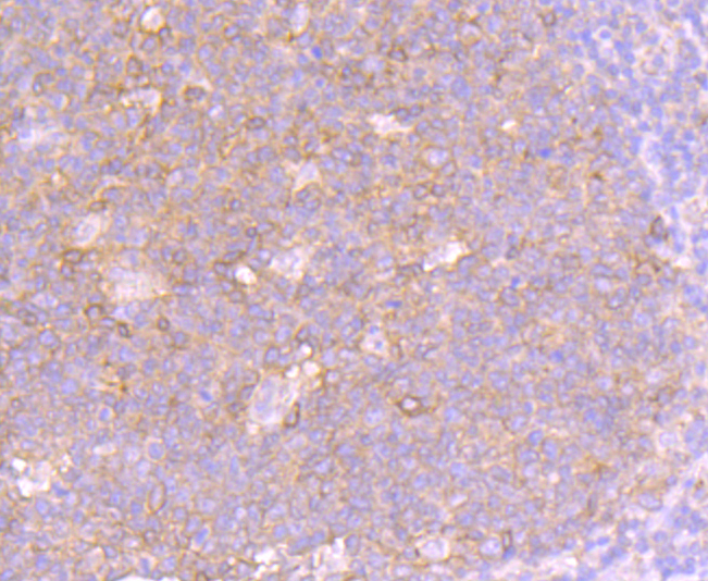
Immunohistochemical analysis of paraffin-embedded human tonsil tissue using anti-Fas antibody. Counter stained with hematoxylin.
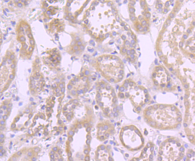
Immunohistochemical analysis of paraffin-embedded human liver cancer tissue using anti-Fas antibody. Counter stained with hematoxylin.
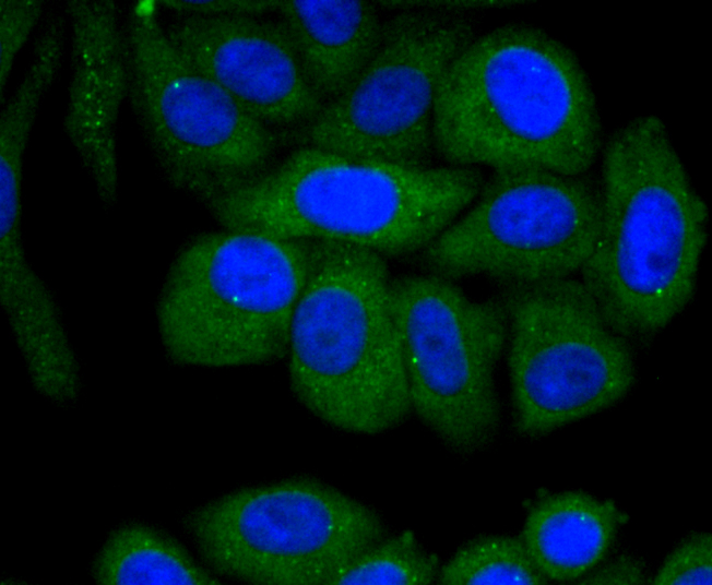
ICC staining Fas in HepG2 cells (green). The nuclear counter stain is DAPI (blue). Cells were fixed in paraformaldehyde, permeabilised with 0.25% Triton X100/PBS.
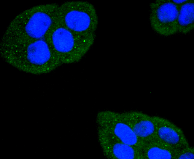
ICC staining Fas in Hela cells (green). The nuclear counter stain is DAPI (blue). Cells were fixed in paraformaldehyde, permeabilised with 0.25% Triton X100/PBS.
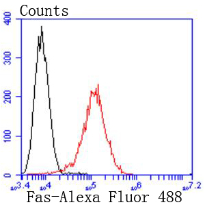
Flow cytometric analysis of Raji cells with Fas antibody at 1/50 dilution (red) compared with an unlabelled control (cells without incubation with primary antibody; black). Alexa Fluor 488-conjugated goat anti rabbit IgG was used as the secondary antibody.
Cytotoxic T lymphocyte (CTL)-mediated cytotoxicity constitutes an important component of specific effector mechanisms in immuno-surveillance against virus-infected or transformed cells. Two mechanisms appear to account for this activity, one of which is the perforin-based process. Independently, a FAS-based mechanism involves the transducing molecule FAS (also designated APO-1) and its ligand (FAS-L). The human FAS protein is a cell surface glycoprotein that belongs to a family of receptors that includes CD40, nerve growth factor receptors and tumor necrosis factor receptors. The FAS antigen is expressed on a broad range of lymphoid cell lines, certain of which undergo apoptosis in response to treatment with antibody to FAS. These findings strongly imply that targeted cell death is potentially mediated by the intercellular interactions of FAS with its ligand or effectors, and that FAS may be critically involved in CTL-mediated cytotoxicity.
If you have published an article using product 49307, please notify us so that we can cite your literature.


 Yes
Yes


