Product Detail
Product NamePDIA6 Rabbit mAb
Clone No.JF0974
Host SpeciesRecombinant Rabbit
Clonality Monoclonal
PurificationProA affinity purified
ApplicationsWB, ICC/IF, FC
Species ReactivityHu, Ms, Rt
Immunogen Descrecombinant protein
ConjugateUnconjugated
Other NamesEndoplasmic reticulum protein 5 antibody ER protein 5 antibody ERp5 antibody P5 antibody Pdia6 antibody PDIA6_HUMAN antibody Protein disulfide isomerase A6 antibody Protein disulfide isomerase associated 6 antibody Protein disulfide isomerase family A member 6 antibody Protein disulfide isomerase P5 antibody Protein disulfide isomerase related protein antibody Protein disulfide-isomerase A6 antibody Thioredoxin domain containing 7 (protein disulfide isomerase) antibody Thioredoxin domain containing protein 7 antibody Thioredoxin domain-containing protein 7 antibody TXNDC7 antibody
Accession NoSwiss-Prot#:Q15084
Uniprot
Q15084
Gene ID
10130;
Calculated MW48 kDa
Formulation1*TBS (pH7.4), 1%BSA, 40%Glycerol. Preservative: 0.05% Sodium Azide.
StorageStore at -20˚C
Application Details
WB: 1:1,000-1:2,000
ICC: 1:100-1:500
FC: 1:50-1:100
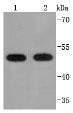
Western blot analysis of PDIA6 on different lysates using anti-PDIA6 antibody at 1/1,000 dilution. Positive control: Lane 1: HepG2 Lane 2: K562
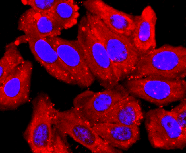
ICC staining PDIA6 in Hela cells (red). The nuclear counter stain is DAPI (blue). Cells were fixed in paraformaldehyde, permeabilised with 0.25% Triton X100/PBS.
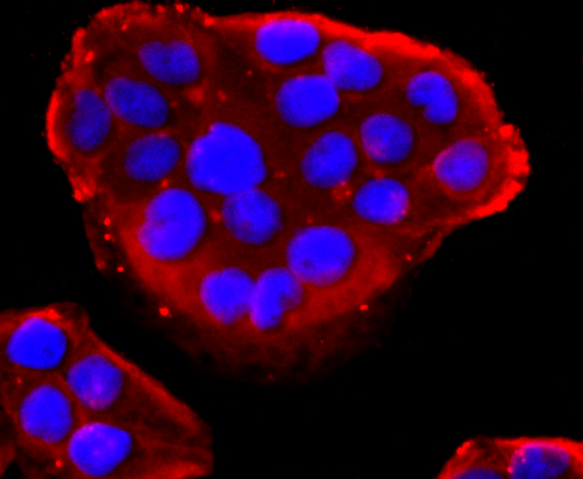
ICC staining PDIA6 in HepG2 cells (red). The nuclear counter stain is DAPI (blue). Cells were fixed in paraformaldehyde, permeabilised with 0.25% Triton X100/PBS.
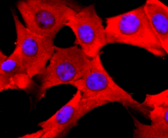
ICC staining PDIA6 in NIH/3T3 cells (green). The nuclear counter stain is DAPI (blue). Cells were fixed in paraformaldehyde, permeabilised with 0.25% Triton X100/PBS.
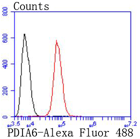
Flow cytometric analysis of Hela cells with PDIA6 antibody at 1/50 dilution (red) compared with an unlabelled control (cells without incubation with primary antibody; black). Alexa Fluor 488-conjugated goat anti rabbit IgG was used as the secondary antibody.
Endoplasmic reticulum proteins (ERps) are widely expressed proteins that localize to the ER and may act as proteases, protein disulfide isomerases, thiol-disulfide oxidases or phospholipases. ERp5, also known as PDIA6 (protein disulfide isomerase family A, member 6) or TXNDC7 is a 440 amino acid protein that contains two thioredoxin domains and belongs to the protein disulfide isomerase family. Localized to the melanosome, as well as to the lumen of the endoplasmic reticulum, ERp5 functions to catalyze the rearrangement of disulfide bonds in a variety of different proteins. Via its catalytic activity, ERp5 is able to reduce the disulfide bond that binds MICA to tumor cells, thereby releasing MICA and reducing the rate of tumor expansion. Multiple isoforms of ERp5 exist due to alternative splicing events.
If you have published an article using product 49340, please notify us so that we can cite your literature.


 Yes
Yes


