Welcome to Sabbiotech!     
Email:info@signalwayantibody.com
Location: Home > Recombinant Rabbit Monoclonal Antibodies > NCAM Rabbit mAb

NCAM Rabbit mAb#49357

Availability: Yes
Total Amount:$
Product Detail

Product NameNCAM Rabbit mAb

Clone No.SR4538

Host SpeciesRecombinant Rabbit

Clonality Monoclonal

PurificationProA affinity purified

ApplicationsWB, ICC/IF, IHC, IP, FC

Species ReactivityHu, Ms, Zebrafish

Immunogen Descrecombinant protein

ConjugateUnconjugated

Other Namesantigen MSK39 identified by monoclonal antibody
5.1H11 antibody
antigen recognized by monoclonal antibody
5.1H11 antibody
CD56 antibody
cell adhesion molecule, neural, 1 antibody
MSK 39 antibody
MSK39 antibody
N-CAM-1 antibody
NCAM 1 antibody
NCAM antibody
NCAM C antibody
NCAM-1 antibody
NCAM1 antibody
NCAM1_HUMAN antibody
NCAMC antibody
Neural cell adhesion molecule 1 antibody
Neural cell adhesion molecule NCAM antibody
OTTHUMP00000235666 antibody

Accession NoSwiss-Prot#:P13591

Uniprot P13591

Gene ID 4684;

Calculated MW100-150 kDa

Formulation1*TBS (pH7.4), 1%BSA, 40%Glycerol. Preservative: 0.05% Sodium Azide.

StorageStore at -20˚C

Application Details
WB: 1:1,000-5,000
IHC: 1:50-1:200
ICC: 1:50-1:200

FC: 1:50-1:100
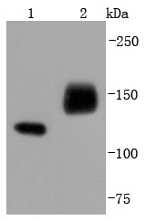
Western blot analysis of NCAM on different lysates using anti-NCAM antibody at 1/1,000 dilution. Positive control:
Lane 1: SH-SY-5Y
Lane 2: Human brain
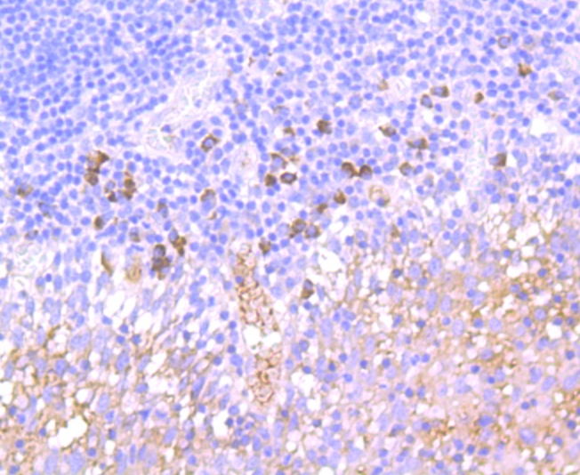
Immunohistochemical analysis of paraffin-embedded human tonsil tissue using anti-NCAM antibody. Counter stained with hematoxylin.
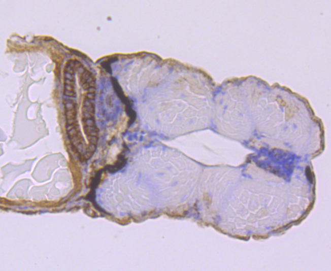
Immunohistochemical analysis of paraffin-embedded zebrafish kidney tissue using anti-NCAM antibody. Counter stained with hematoxylin.
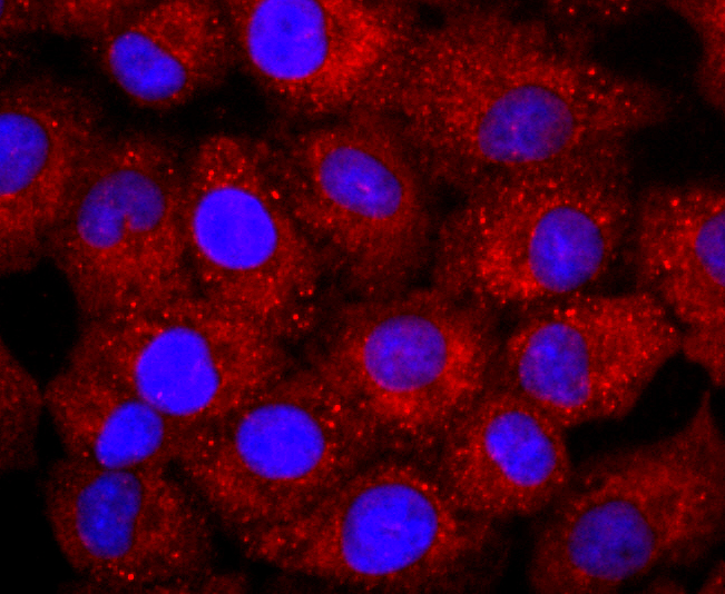
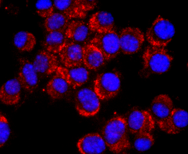
ICC staining NCAM in A549 cells (red). The nuclear counter stain is DAPI (blue). Cells were fixed in paraformaldehyde, permeabilised with 0.25% Triton X100/PBS.
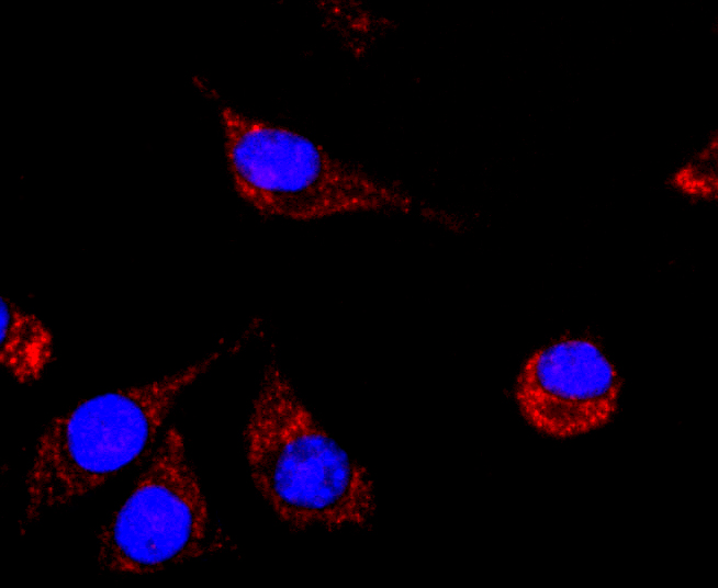
ICC staining NCAM in N2A cells (red). The nuclear counter stain is DAPI (blue). Cells were fixed in paraformaldehyde, permeabilised with 0.25% Triton X100/PBS.
ICC staining NCAM in SH-SY-5Y cells (red). The nuclear counter stain is DAPI (blue). Cells were fixed in paraformaldehyde, permeabilised with 0.25% Triton X100/PBS.
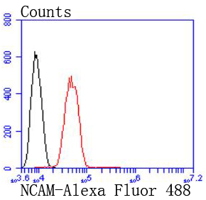
Flow cytometric analysis of SH-SY-5Y cells with NCAM antibody at 1/50 dilution (red) compared with an unlabelled control (cells without incubation with primary antibody; black). Alexa Fluor 488-conjugated goat anti rabbit IgG was used as the secondary antibody.
Neural cell adhesion molecules (NCAMs) are a family of closely related cell surface glycoproteins involved in cell to cell interactions during growth and thought to play an important role in embryogenesis and development. The expression of these molecules is widespread in all three germ layers during embryogenesis, but is more restrictive in adult tissues. NCAM expression is observed in a variety of human tumors including neuroblastomas, rhabdo-myosarcomas, Wilms' tumor, Ewing's sarcoma and some primitive myeloid malignancies. Multiple isoforms of NCAM have been reported in both mouse and human brain tissue. In humans, NCAMs arise from differential splicing and use of alternative polyadenylation sites of a single gene mapping to 11q23.

If you have published an article using product 49357, please notify us so that we can cite your literature.

NOTE

Application

  • WBWestern Blotting
  • IHCImmunohistochemistry
  • IFImmunofluorescence
  • ICCImmunocytochemistry
  • FCFlow Cytometry
  • IPImmunoprecipitation
  • EELISA
  • DBDot Blotting
  • ChIPChromatin Immunoprecipitation
  • GICAGold Immunochromatography Assay
  • NCNegative Control

Species Reactivity

  • HuHuman
  • MsMouse
  • RtRat
  • DmDrosophila melanogaster
  • CCaenorhabditis elegans
  • MkMonkey
  • RbRabbit
  • BBovine
  • DDog
  • PPig
  • HmHamster
  • ChHmChinese Hamster
  • ChkChicken
  • ShpSheep
;