Location:
Home
>
Recombinant Rabbit Monoclonal Antibodies > EDG1 Rabbit mAb
EDG1 Rabbit mAb#49440

 Yes
Yes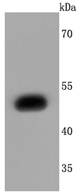
Western blot analysis of EDG1 on SH-SY5Y cells lysates using anti-EDG1 antibody at 1/1,000 dilution.
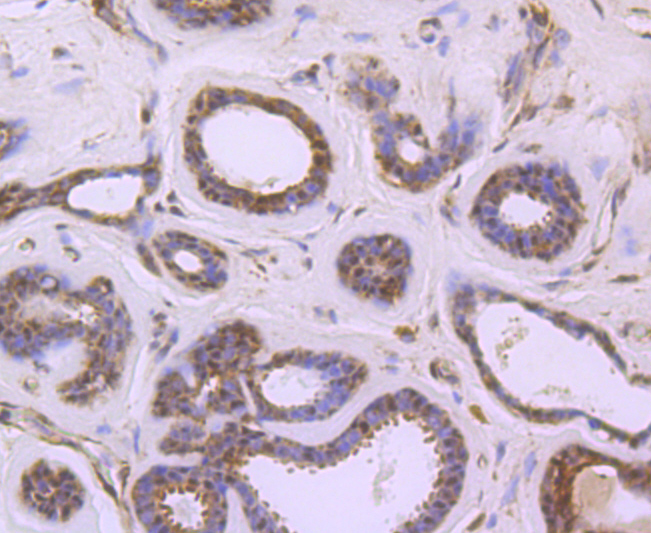
Immunohistochemical analysis of paraffin-embedded human breast carcinoma tissue using anti-EDG1 antibody. Counter stained with hematoxylin.
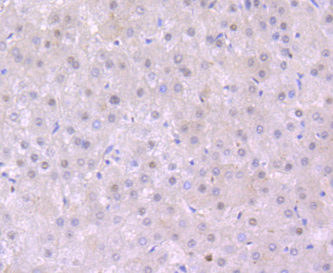
Immunohistochemical analysis of paraffin-embedded human liver tissue using anti-EDG1 antibody. Counter stained with hematoxylin.
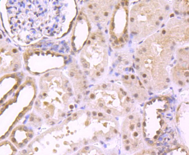
Immunohistochemical analysis of paraffin-embedded human kidney tissue using anti-EDG1 antibody. Counter stained with hematoxylin.
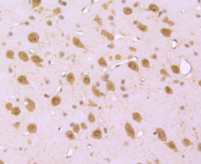
Immunohistochemical analysis of paraffin-embedded mouse brain tissue using anti-EDG1 antibody. Counter stained with hematoxylin.

Immunohistochemical analysis of paraffin-embedded mouse herat tissue using anti-EDG1 antibody. Counter stained with hematoxylin.
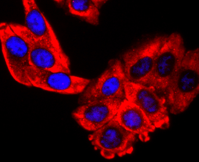
ICC staining EDG1 in HepG2 cells (red). The nuclear counter stain is DAPI (blue). Cells were fixed in paraformaldehyde, permeabilised with 0.25% Triton X100/PBS.
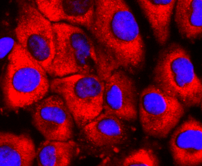
ICC staining EDG1 in HUVEC cells (red). The nuclear counter stain is DAPI (blue). Cells were fixed in paraformaldehyde, permeabilised with 0.25% Triton X100/PBS.
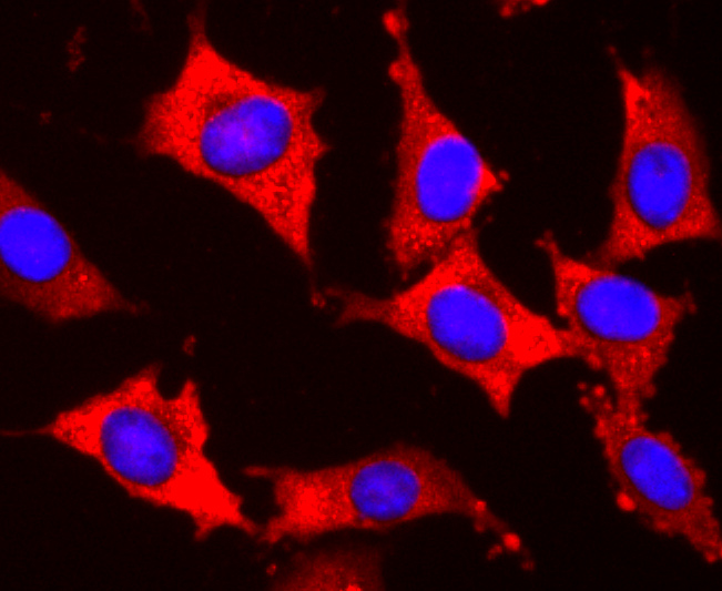
ICC staining EDG1 in SH-SY5Y cells (red). The nuclear counter stain is DAPI (blue). Cells were fixed in paraformaldehyde, permeabilised with 0.25% Triton X100/PBS.
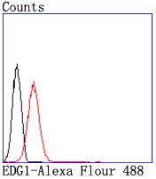
Flow cytometric analysis of Jurkat cells with EDG1 antibody at 1/50 dilution (red) compared with an unlabelled control (cells without incubation with primary antibody; black). Alexa Fluor 488-conjugated goat anti rabbit IgG was used as the secondary antibody.
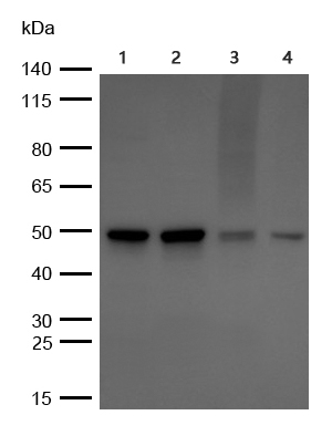
All lanes : EDG1 Rabbit mAb at 1/1000 dilution
Lane 1 :HUVEC whole cell lysates
Lane 2 :JK whole cell lysates
Lane 3 :HT29 whole cell lysates
Lane 4 :Rat brain lysates
Lysates at 20 µg per lane.
Secondary
All lanes : Goat Anti-Rabbit IgG H&L (HRP) at 1/10000 dilution
Predicted band size: 43 kDa
Observed band size: 43-50 kDa
Exposure time: 5 seconds
NOTE
Application
- WBWestern Blotting
- IHCImmunohistochemistry
- IFImmunofluorescence
- ICCImmunocytochemistry
- FCFlow Cytometry
- IPImmunoprecipitation
- EELISA
- DBDot Blotting
- ChIPChromatin Immunoprecipitation
- GICAGold Immunochromatography Assay
- NCNegative Control
Species Reactivity
- HuHuman
- MsMouse
- RtRat
- DmDrosophila melanogaster
- CCaenorhabditis elegans
- MkMonkey
- RbRabbit
- BBovine
- DDog
- PPig
- HmHamster
- ChHmChinese Hamster
- ChkChicken
- ShpSheep


