
 Yes
Yes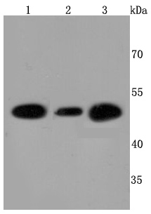
Western blot analysis of ATPB on different cells lysates using anti-ATPB antibody at 1/500 dilution. Positive control: Line1: Hela Line2: HepG2 Line3: 293T
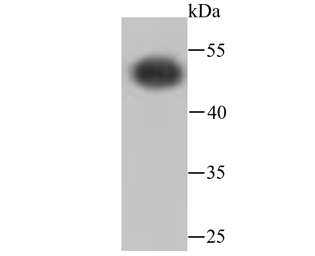
Western blot analysis of ATPB on Zebrafish cells lysates using anti-ATPB antibody at 1/500 dilution.
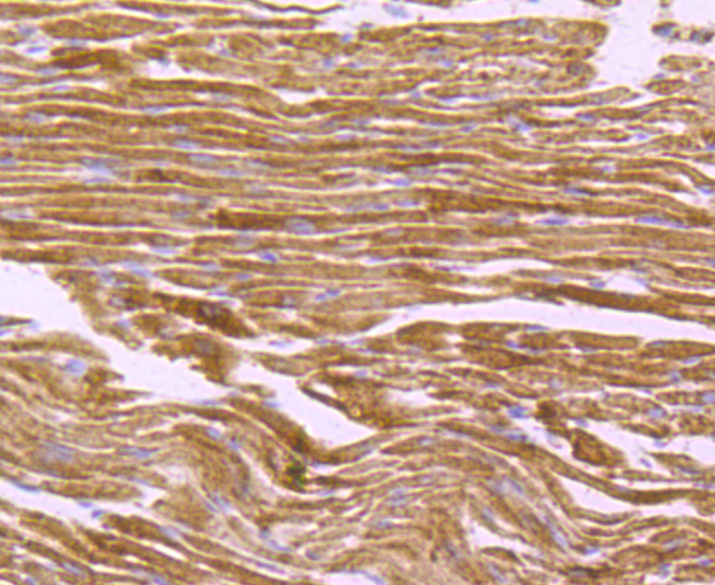
Immunohistochemical analysis of paraffin-embedded mouse heart tissue using anti-ATPB antibody. Counter stained with hematoxylin.
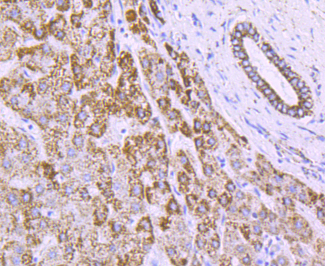
Immunohistochemical analysis of paraffin-embedded human liver tissue using anti-ATPB antibody. Counter stained with hematoxylin.
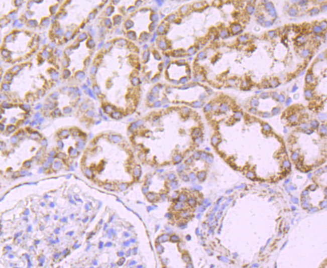
Immunohistochemical analysis of paraffin-embedded human kidney tissue using anti-ATPB antibody. Counter stained with hematoxylin.
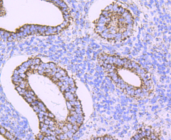
Immunohistochemical analysis of paraffin-embedded huaman uterus tissue using anti-ATPB antibody. Counter stained with hematoxylin.
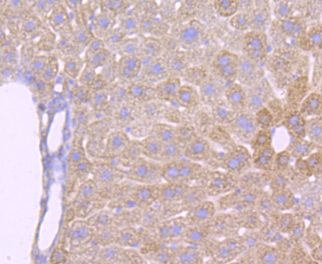
Immunohistochemical analysis of paraffin-embedded mouse liver tissue using anti-ATPB antibody. Counter stained with hematoxylin.
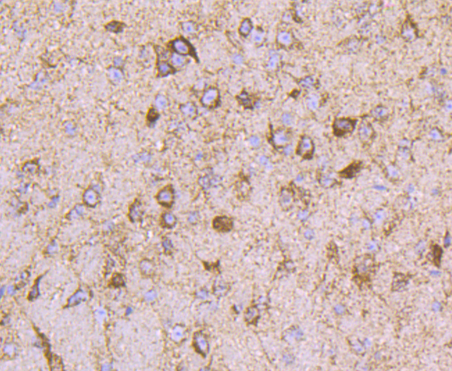
Immunohistochemical analysis of paraffin-embedded mouse brain tissue using anti-ATPB antibody. Counter stained with hematoxylin.
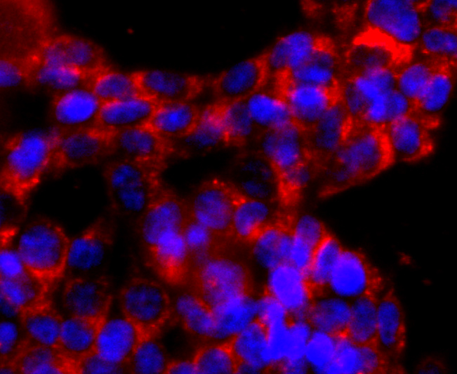
ICC staining ATPB in 293T cells (red). The nuclear counter stain is DAPI (blue). Cells were fixed in paraformaldehyde, permeabilised with 0.25% Triton X100/PBS.
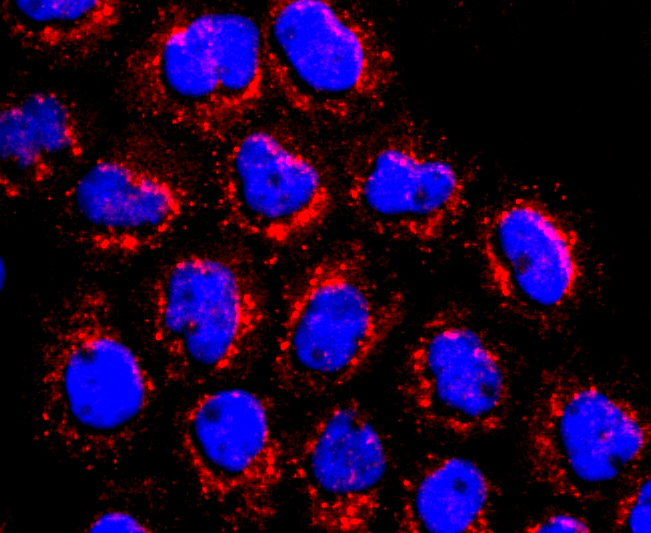
ICC staining ATPB in A431 cells (red). The nuclear counter stain is DAPI (blue). Cells were fixed in paraformaldehyde, permeabilised with 0.25% Triton X100/PBS.
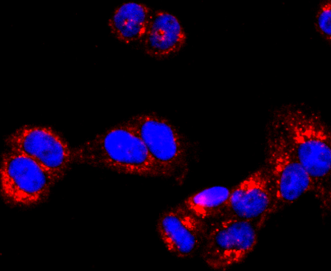
ICC staining ATPB in Hela cells (red). The nuclear counter stain is DAPI (blue). Cells were fixed in paraformaldehyde, permeabilised with 0.25% Triton X100/PBS.
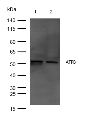
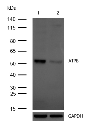
All lanes : ATPB Rabbit mAb at 1/1k dilutionLane 1 : Rat spleen lysates whole cell lysatesLane 2 : Mouse spleen lysates whole cell lysatesLysates/proteins at 20 µg per lane.SecondaryAll lanes : Goat Anti-Rabbit IgG H&L (HRP) at 1/20000 dilutionPredicted band size: 57 kDa Observed band size: 53 kDaExposure time: 6 seconds
All lanes : ATPB Rabbit mAb at 1/1k dilution Lane 1 : Wild-type HAP1 cell lysate Lane 2 : ATPB knockdown HAP1 cell lysate Lysates/proteins at 20 µg per lane.
NOTE
Application
- WBWestern Blotting
- IHCImmunohistochemistry
- IFImmunofluorescence
- ICCImmunocytochemistry
- FCFlow Cytometry
- IPImmunoprecipitation
- EELISA
- DBDot Blotting
- ChIPChromatin Immunoprecipitation
- GICAGold Immunochromatography Assay
- NCNegative Control
Species Reactivity
- HuHuman
- MsMouse
- RtRat
- DmDrosophila melanogaster
- CCaenorhabditis elegans
- MkMonkey
- RbRabbit
- BBovine
- DDog
- PPig
- HmHamster
- ChHmChinese Hamster
- ChkChicken
- ShpSheep


