Product Detail
Product NameFGFR3 Rabbit mAb
Clone No.JM81-10
Host SpeciesRecombinant Rabbit
Clonality Monoclonal
PurificationProA affinity purified
ApplicationsWB, ICC/IF, IHC, FC
Species ReactivityHu
Immunogen Descrecombinant protein
ConjugateUnconjugated
Other NamesACH antibody
CD 333 antibody
CD333 antibody
CD333 antigen antibody
CEK 2 antibody
CEK2 antibody
FGFR 3 antibody
FGFR-3 antibody
FGFR3 antibody
FGFR3_HUMAN antibody
Fibroblast growth factor receptor 3 (achondroplasia thanatophoric dwarfism) antibody
Fibroblast growth factor receptor 3 antibody
Heparin binding growth factor receptor antibody
HSFGFR3EX antibody
Hydroxyaryl protein kinase antibody
JTK 4 antibody
JTK4 antibody
MFR 3 antibody
SAM 3 antibody
Tyrosine kinase JTK 4 antibody
Tyrosine kinase JTK4 antibody
Z FGFR 3 antibody
Accession NoSwiss-Prot#:P22607
Uniprot
P22607
Gene ID
2261;
Calculated MW100 kDa
Formulation1*TBS (pH7.4), 1%BSA, 40%Glycerol. Preservative: 0.05% Sodium Azide.
StorageStore at -20˚C
Application Details
WB: 1:1,000-5,000
IHC: 1:50-1:200
ICC: 1:50-1:200
FC:150-1100
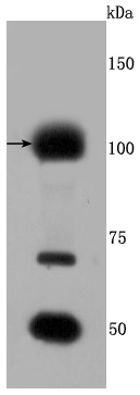
Western blot analysis of FGFR3 on Hela cells lysates using anti-FGFR3 antibody at 1/500 dilution.
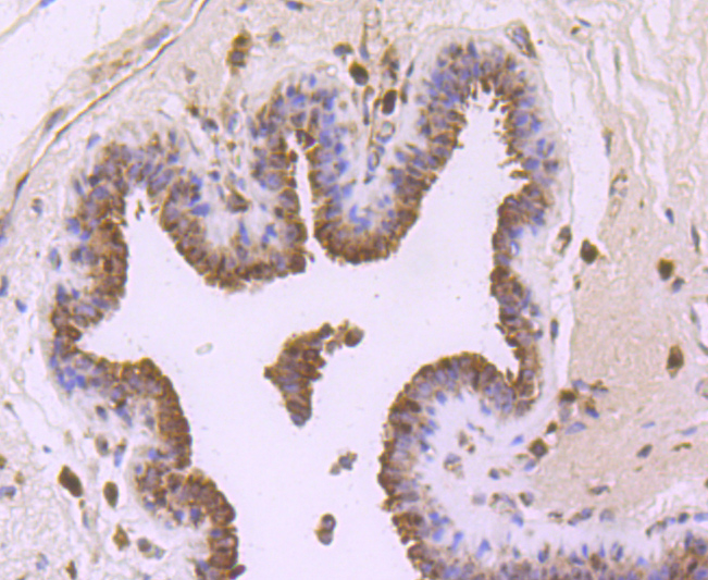
Immunohistochemical analysis of paraffin-embedded human breast cancer tissue using anti-FGFR3 antibody. Counter stained with hematoxylin.
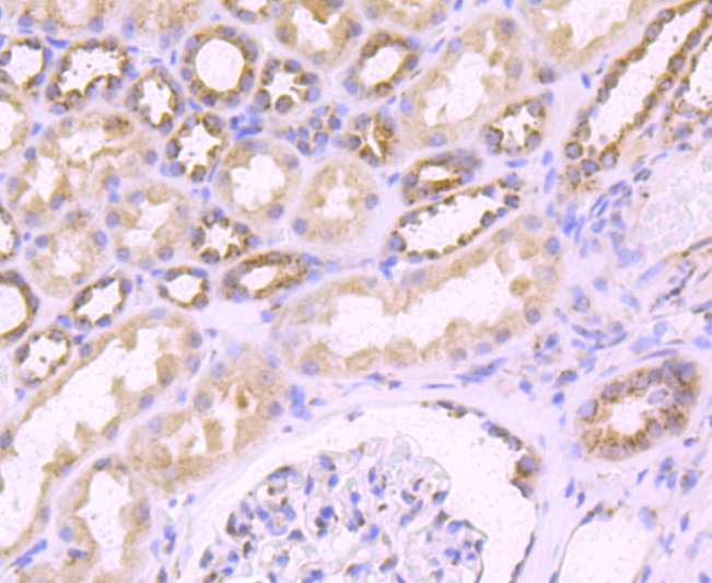
Immunohistochemical analysis of paraffin-embedded human kidney tissue using anti-FGFR3 antibody. Counter stained with hematoxylin.
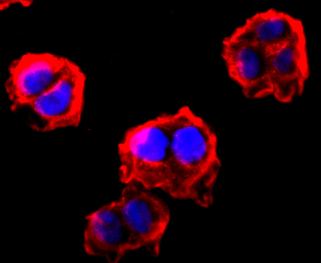
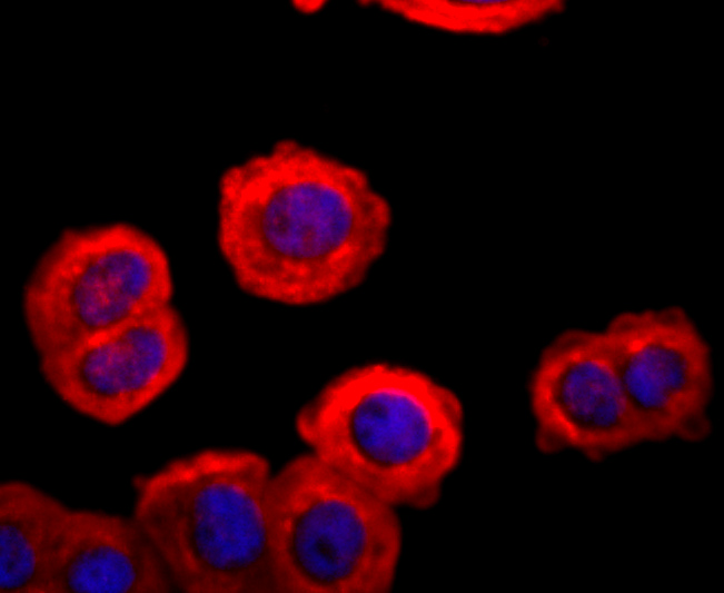
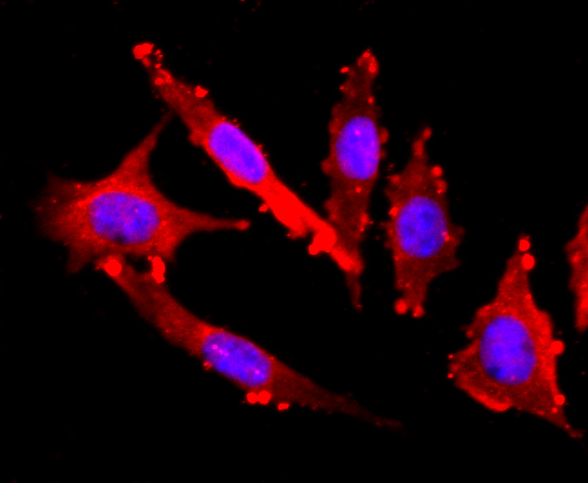
ICC staining FGFR3 in Hela cells (red). The nuclear counter stain is DAPI (blue). Cells were fixed in paraformaldehyde, permeabilised with 0.25% Triton X100/PBS.
ICC staining FGFR3 in MCF-7 cells (red). The nuclear counter stain is DAPI (blue). Cells were fixed in paraformaldehyde, permeabilised with 0.25% Triton X100/PBS.
ICC staining FGFR3 in SH-SY5Y cells (red). The nuclear counter stain is DAPI (blue). Cells were fixed in paraformaldehyde, permeabilised with 0.25% Triton X100/PBS.
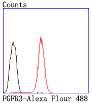
Flow cytometric analysis of HepG2 cells with FGFR3 antibody at 1/50 dilution (red) compared with an unlabelled control (cells without incubation with primary antibody; black). Alexa Fluor 488-conjugated goat anti rabbit IgG was used as the secondary� antibody.
Acidic and basic fibroblast growth factors (FGFs) are members of a family of multifunctional polypeptide growth factors that stimulate proliferation of cells of mesenchymal, epithelial and neuroectodermal origin. Like other growth factors, FGFs act by binding and activating specific cell surface receptors. These include the Flg receptor or FGFR-1, the Bek receptor or FGFR-2, FGFR-3, FGFR-4, FGFR-5 and FGFR-6. These receptors usually contain an extracellular ligand-binding region containing three immunoglobulin-like domains, a transmembrane domain and a cytoplasmic tyrosine kinase domain. The gene encoding human FGFR-3 maps to chromosome 4p16 and is alternatively spliced to produce three isoforms that are expressed in brain, kidney and testis. Defects in FGFR-3 are associated with several diseases, including Crouzon syndrome, achondroplasia, thanatophoric dysplasia, craniosynostosis adelaide type and hypochondroplasia. Mutations in FGFR-3 are also a cause of some bladder and cervical cancers.
If you have published an article using product 49449, please notify us so that we can cite your literature.


 Yes
Yes


