Product Detail
Product NameTNF Receptor II Rabbit mAb
Clone No.SR4542
Host SpeciesRecombinant Rabbit
Clonality Monoclonal
PurificationProA affinity purified
ApplicationsWB, ICC/IF, IHC, FC, IP
Species ReactivityHu, Ms, Rt
Immunogen Descrecombinant protein
ConjugateUnconjugated
Other NamesCD120b antibody
p75 antibody
p75 TNF receptor antibody
p75TNFR antibody
p80 TNF alpha receptor antibody
p80 TNF-alpha receptor antibody
Soluble TNFR1B variant 1 antibody
TBP-2 antibody
TBPII antibody
TNF R II antibody
TNF R2 antibody
TNF R75 antibody
TNF-R2 antibody
TNF-RII antibody
TNFBR antibody
TNFR-II antibody
TNFR1B antibody
TNFR2 antibody
TNFR80 antibody
TNFRII antibody
Tnfrsf1b antibody
TNR1B_HUMAN antibody
Tumor necrosis factor beta receptor antibody
Tumor necrosis factor receptor 2 antibody
Tumor necrosis factor receptor superfamily member 1B antibody
Tumor necrosis factor receptor type II antibody
Tumor necrosis factor-binding protein 2 antibody
Accession NoSwiss-Prot#:P20333
Uniprot
P20333
Gene ID
7133;
Calculated MW73 kDa
Formulation1*TBS (pH7.4), 1%BSA, 40%Glycerol. Preservative: 0.05% Sodium Azide.
StorageStore at -20˚C
Application Details
WB: 1:500-1:2,000
IHC: 1:50-1:200
ICC: 1:50-1:200
IP: 1:10-1:50
FC: 1:50-1:100
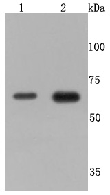
Western blot analysis of TNF Receptor II on different cells lysates using anti-TNF Receptor II antibody at 1/500 dilution. Positive control��
Line 1: MCF-7
Line 2: Jurkat
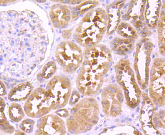
Immunohistochemical analysis of paraffin-embedded human kidney tissue using anti-TNF Receptor II antibody. Counter stained with hematoxylin.
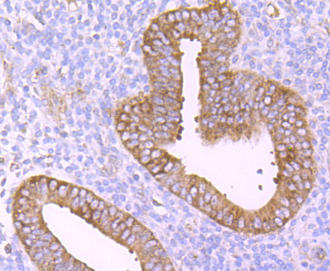
Immunohistochemical analysis of paraffin-embedded human uterus tissue using anti-TNF Receptor II antibody. Counter stained with hematoxylin.
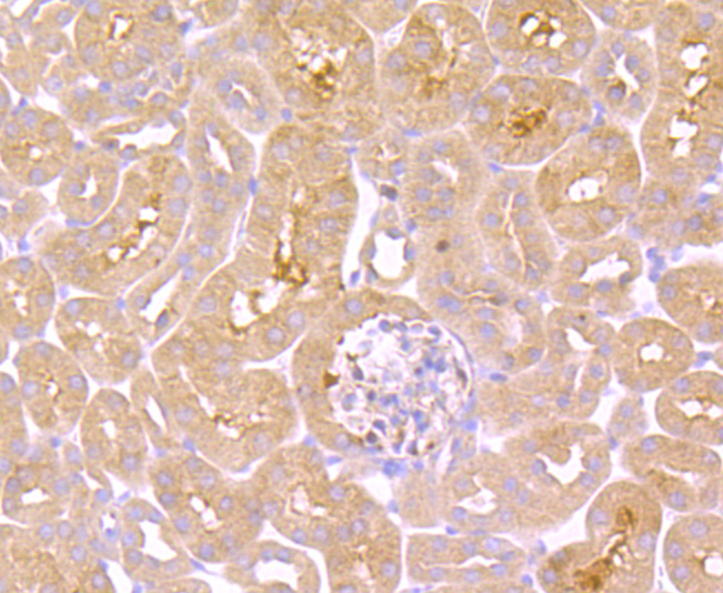
Immunohistochemical analysis of paraffin-embedded mouse kidney tissue anti-TNF Receptor II antibody. Counter stained with hematoxylin.
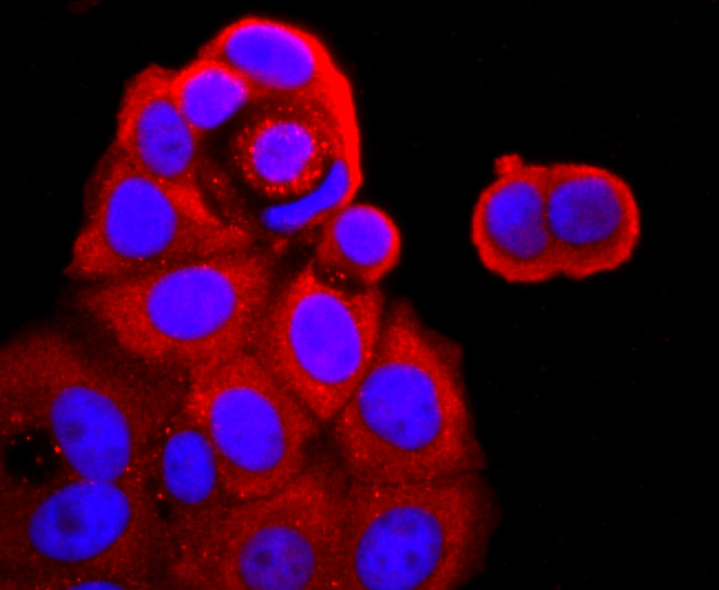
ICC staining TNF Receptor II in MCF-7 cells (red). The nuclear counter stain is DAPI (blue). Cells were fixed in paraformaldehyde, permeabilised with 0.25% Triton X100/PBS.
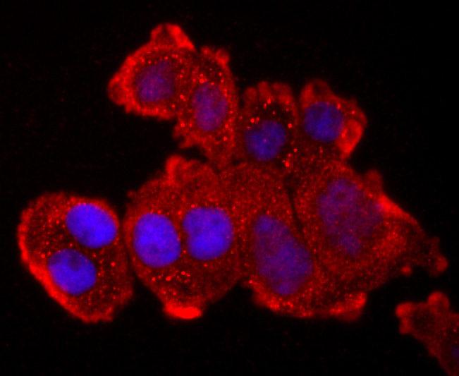
ICC staining TNF Receptor II in Hela cells (red). The nuclear counter stain is DAPI (blue). Cells were fixed in paraformaldehyde, permeabilised with 0.25% Triton X100/PBS.
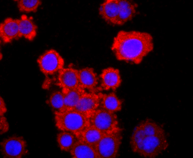
ICC staining TNF Receptor II in SW480 cells (red). The nuclear counter stain is DAPI (blue). Cells were fixed in paraformaldehyde, permeabilised with 0.25% Triton X100/PBS.
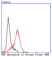
Flow cytometric analysis of HL-60 cells with TNF Receptor II antibody at 1/50 dilution (red) compared with an unlabelled control (cells without incubation with primary antibody; black). Alexa Fluor 488-conjugated goat anti rabbit IgG was used as the secondary antibody.
Tumor necrosis factor (TNF) is a pleiotropic cytokine whose function is mediated through two distinct cell surface receptors. These receptors, designated TNF-R1 and TNF-R2, are expressed on most cell types. The majority of TNF functions are primarily mediated through TNF-R1, while signaling through TNF-R2 occurs less extensively and is confined to cells of the immune system. Both of these proteins belong to the growing TNF and nerve growth factor (NGF) receptor superfamily, which includes FAS, CD30, CD27 and CD40. The members of this superfamily are type I membrane proteins that share sequence homology confined to the extracellular region. TNF-R1 shares a motif termed the "death domain" with FAS and three structurally unrelated signaling proteins, TRADD, FADD and RIP (1,3-8). This death domain is required for transduction of the apoptotic signal.
If you have published an article using product 49476, please notify us so that we can cite your literature.


 Yes
Yes


