Product Detail
Product NameS100 Rabbit mAb
Clone No.JM51-31
Host SpeciesRecombinant Rabbit
Clonality Monoclonal
PurificationProA affinity purified
ApplicationsWB, IP, IHC, FC
Species ReactivityHu, Ms
Immunogen Descrecombinant protein
ConjugateUnconjugated
Other NamesNEF antibody
Protein S100 B antibody
Protein S100-B antibody
S 100 calcium binding protein beta chain antibody
S 100 protein beta chain antibody
S-100 protein beta chain antibody
S-100 protein subunit beta antibody
S100 antibody
S100 calcium binding protein beta (neural) antibody
S100 calcium-binding protein B antibody
S100 protein beta chain antibody
S100B antibody
S100B_HUMAN antibody
S100beta antibody
Accession NoSwiss-Prot#:P23297
Uniprot
P23297
Gene ID
6271;
Calculated MW11 kDa
Formulation1*TBS (pH7.4), 1%BSA, 40%Glycerol. Preservative: 0.05% Sodium Azide.
StorageStore at -20˚C
Application Details
WB: 1:500-1:2,000
IHC: 1:50-1:200
IP: 1:10-1:50
FC: 1:50-1:100
Western blot analysis of S100 on mouse heart cells lysates using anti-S100 antibody at 1/500 dilution.
Immunohistochemical analysis of paraffin-embedded human tonsil tissue using anti- S100 antibody. Counter stained with hematoxylin.
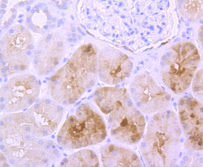
Immunohistochemical analysis of paraffin-embedded human kidney tissue using anti- S100 antibody. Counter stained with hematoxylin.
Immunohistochemical analysis of paraffin-embedded mouse brain tissue using anti- S100 antibody. Counter stained with hematoxylin.
Immunohistochemical analysis of paraffin-embedded mouse spinal cord tissue using anti-S100 antibody. Counter stained with hematoxylin.
Flow cytometric analysis of SH-SY5Y cells with S100 antibody at 1/50 dilution (red) compared with an unlabelled control (cells without incubation with primary antibody; black). Alexa Fluor 488-conjugated goat anti rabbit IgG was used as the secondary antibody.
The family of EF-hand type Ca2+-binding proteins includes calbindin (previously designated vitamin D-dependent Ca2+-binding protein), S-100 α and β, calgranulins A (also designated MRP8), B (also designated MRP14) and C (S-100 like proteins), and the parvalbumin family members, including parvalbumin α and parvalbumin β (also designated oncomodulin). The S-100 protein is involved in the regulation of cellular processes such as cell cycle progression and differentiation. Research also indicates that the S-100 protein may function in the activation of Ca2+ induced Ca2+ release, inhibition of microtubule assembly and inhibition of protein kinase C mediated phosphorylation. Two S-100 subunits, sharing 60% sequence identity, have been described as S-100 α chain and S-100 β chain. Three S-100 dimeric forms have been characterized, differing in their subunit composition of either two α chains, two β chains or one α and one β chain. S-100 localizes to the cytoplasm and nuclei of astrocytes, Schwann's cells, ependymomas and astrogliomas. S-100 is also detected in almost all benign naevi, malignant melanocytic tumours and in Langerhans cells in the skin. Calbindin, S-100 proteins and parvalbumin proteins are each expressed in neural tissues. In addition, S-100 α and β are present in a variety of other tissues, and calbindin is present in intestine and kidney.
If you have published an article using product 49485, please notify us so that we can cite your literature.


 Yes
Yes


