Welcome to Sabbiotech!     
Email:info@signalwayantibody.com
Location: Home > Recombinant Rabbit Monoclonal Antibodies > 53BP1 Rabbit mAb

53BP1 Rabbit mAb#49517

Availability: Yes
Total Amount:$
Product Detail

Product Name53BP1 Rabbit mAb

Clone No.JA66-11

Host SpeciesRecombinant Rabbit

Clonality Monoclonal

PurificationProA affinity purified

ApplicationsWB, ICC/IF, IHC, FC

Species ReactivityHu, Ms, Rt

Immunogen Descrecombinant protein

ConjugateUnconjugated

Other Names53 BP1 antibody
53BP1 antibody
FLJ41424 antibody
MGC138366 antibody
p202 antibody
p53 binding protein 1 antibody
p53 BP1 antibody
p53-binding protein 1 antibody
p53BP1 antibody
TP53 BP1 antibody
TP53B_HUMAN antibody
Tp53bp1 antibody
TRP53 BP1 antibody
Tumor protein 53 binding protein 1 antibody
Tumor protein p53 binding protein 1 antibody
Tumor suppressor p53 binding protein 1 antibody
Tumor suppressor p53-binding protein 1 antibody

Accession NoSwiss-Prot#:Q12888

Uniprot Q12888

Gene ID 7158;

Calculated MW214 kDa

Formulation1*TBS (pH7.4), 1%BSA, 40%Glycerol. Preservative: 0.05% Sodium Azide.

StorageStore at -20˚C

Application Details
WB: 1:500-1:1,000
IHC: 1:50-1:200
ICC: 1:100-1:500

FC: 1:50-1:100
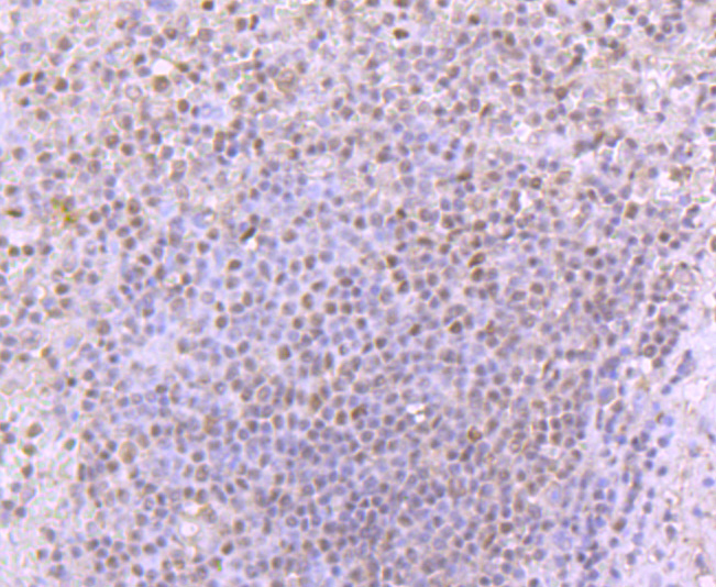
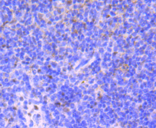
Immunohistochemical analysis of paraffin-embedded human spleen tissue using anti-53BP1 antibody. Counter stained with hematoxylin.
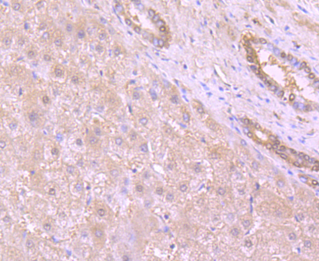
Immunohistochemical analysis of paraffin-embedded human liver tissue using anti-53BP1 antibody. Counter stained with hematoxylin.
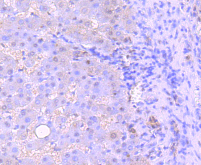
Immunohistochemical analysis of paraffin-embedded human liver cancer tissue using anti-53BP1 antibody. Counter stained with hematoxylin.
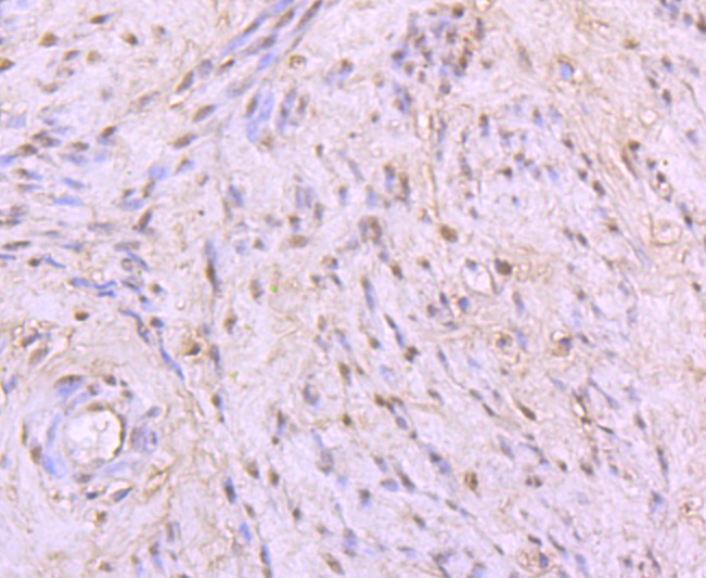
Immunohistochemical analysis of paraffin-embedded human cervix tissue using anti-53BP1 antibody. Counter stained with hematoxylin.
Immunohistochemical analysis of paraffin-embedded mouse spleen tissue using anti-53BP1 antibody. Counter stained with hematoxylin.
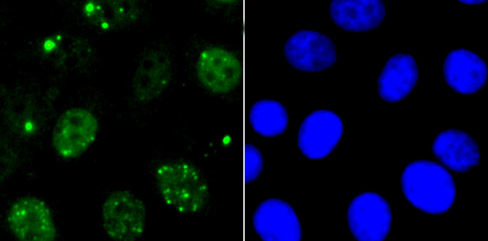
ICC staining 53BP1 in HepG2 cells (green). The nuclear counter stain is DAPI (blue). Cells were fixed in paraformaldehyde, permeabilised with 0.25% Triton X100/PBS.
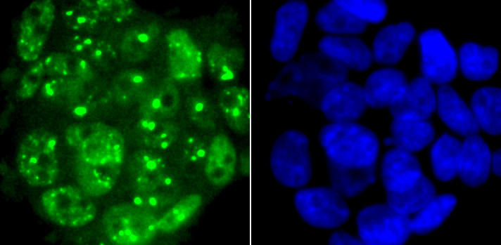
ICC staining 53BP1 in 293T cells (green). The nuclear counter stain is DAPI (blue). Cells were fixed in paraformaldehyde, permeabilised with 0.25% Triton X100/PBS.
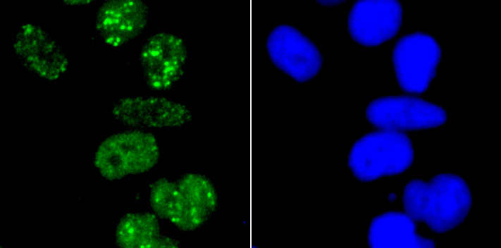
ICC staining 53BP1 in Hela cells (green). The nuclear counter stain is DAPI (blue). Cells were fixed in paraformaldehyde, permeabilised with 0.25% Triton X100/PBS.
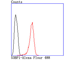
Flow cytometric analysis of Hela cells with 53BP1 antibody at 1/50 dilution (red) compared with an unlabelled control (cells without incubation with primary antibody; black). Alexa Fluor 488-conjugated goat anti rabbit IgG was used as the secondary antibody.
The p53 binding proteins 53BP1 and 53BP2 (Bbp) bind to the central DNA-binding domain of wild type p53, but do not bind mutant p53. The central DNA-binding domain of p53 is required for site-specific DNA binding and is frequently mutated in malignant tumors. Binding of 53BP1 to the L3 loop of p53 and of 53BP2 to the L2 loop of p53 confirms that the loop is dependent on p53 conformation. Site-specific binding also suggests that 53BP1 and 53BP2 are involved in p53-mediated tumor suppression. 53BP1 was isolated from H258 cells and is expressed in Jurkat cells in both the cytoplasm and the nucleus. The N-terminus of 53BP2 is localized to the cytoplasm, while the C-terminus might be localized in the nucleus. 53BP1 promotes cell proliferation by binding to p202, whereas 53BP2 induces cell death by binding to Bcl2 and NFkB p65.

If you have published an article using product 49517, please notify us so that we can cite your literature.

NOTE

Application

  • WBWestern Blotting
  • IHCImmunohistochemistry
  • IFImmunofluorescence
  • ICCImmunocytochemistry
  • FCFlow Cytometry
  • IPImmunoprecipitation
  • EELISA
  • DBDot Blotting
  • ChIPChromatin Immunoprecipitation
  • GICAGold Immunochromatography Assay
  • NCNegative Control

Species Reactivity

  • HuHuman
  • MsMouse
  • RtRat
  • DmDrosophila melanogaster
  • CCaenorhabditis elegans
  • MkMonkey
  • RbRabbit
  • BBovine
  • DDog
  • PPig
  • HmHamster
  • ChHmChinese Hamster
  • ChkChicken
  • ShpSheep
;