Location:
Home
>
Recombinant Rabbit Monoclonal Antibodies > MUC2 Rabbit mAb
MUC2 Rabbit mAb#49518

 Yes
Yes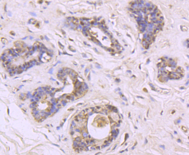
Immunohistochemical analysis of paraffin-embedded human breast cancer tissue using anti-MUC2 antibody. Counter stained with hematoxylin.
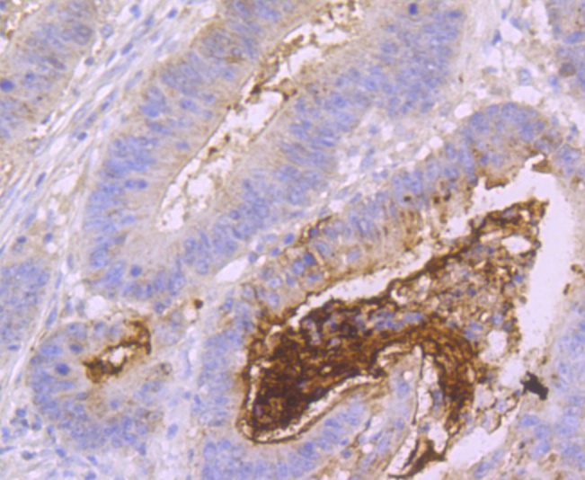
Immunohistochemical analysis of paraffin-embedded human colon cancer tissue using anti-MUC2 antibody. Counter stained with hematoxylin.
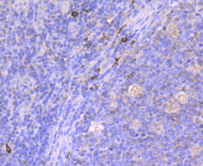
Immunohistochemical analysis of paraffin-embedded human tonsil tissue using anti-MUC2 antibody. Counter stained with hematoxylin.
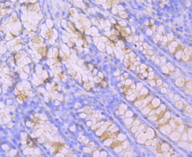
Immunohistochemical analysis of paraffin-embedded rat intestine tissue using anti-MUC2 antibody. Counter stained with hematoxylin.
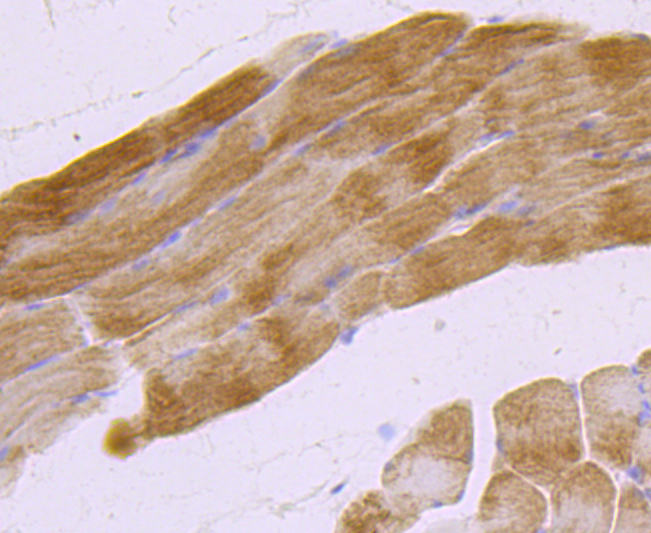
Immunohistochemical analysis of paraffin-embedded rat skeletal muscle tissue using anti-MUC2 antibody. Counter stained with hematoxylin.
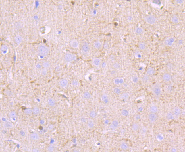
Immunohistochemical analysis of paraffin-embedded rat brain tissue using anti-MUC2 antibody. Counter stained with hematoxylin.
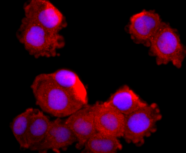
ICC staining MUC2 in Hela cells (red). The nuclear counter stain is DAPI (blue). Cells were fixed in paraformaldehyde, permeabilised with 0.25% Triton X100/PBS.
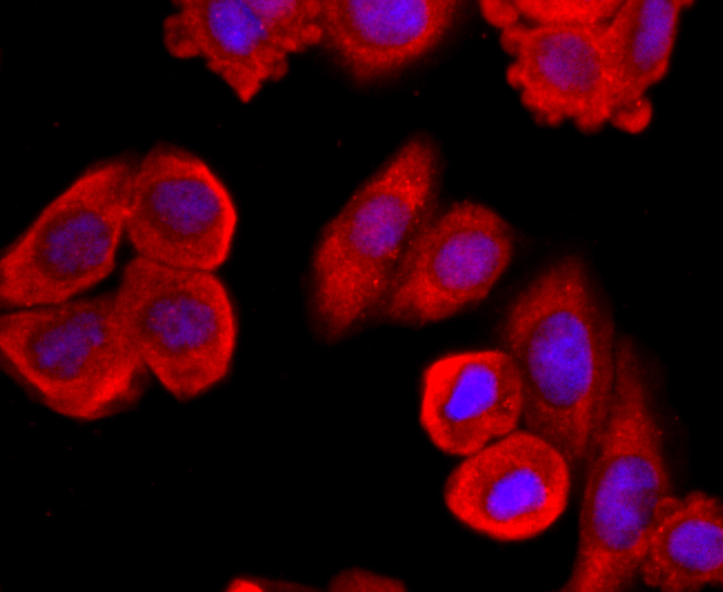
ICC staining MUC2 in HepG2 cells (red). The nuclear counter stain is DAPI (blue). Cells were fixed in paraformaldehyde, permeabilised with 0.25% Triton X100/PBS.
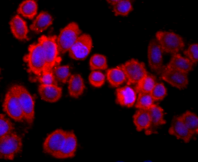
ICC staining MUC2 in SW480 cells (red). The nuclear counter stain is DAPI (blue). Cells were fixed in paraformaldehyde, permeabilised with 0.25% Triton X100/PBS.
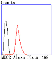
Flow cytometric analysis of Hela cells with MUC2 antibody at 1/50 dilution (red) compared with an unlabelled control (cells without incubation with primary antibody; black). Alexa Fluor 488-conjugated goat anti rabbit IgG was used as the secondary antibody.
NOTE
Application
- WBWestern Blotting
- IHCImmunohistochemistry
- IFImmunofluorescence
- ICCImmunocytochemistry
- FCFlow Cytometry
- IPImmunoprecipitation
- EELISA
- DBDot Blotting
- ChIPChromatin Immunoprecipitation
- GICAGold Immunochromatography Assay
- NCNegative Control
Species Reactivity
- HuHuman
- MsMouse
- RtRat
- DmDrosophila melanogaster
- CCaenorhabditis elegans
- MkMonkey
- RbRabbit
- BBovine
- DDog
- PPig
- HmHamster
- ChHmChinese Hamster
- ChkChicken
- ShpSheep


