Product Detail
Product NameNFAT2 Rabbit mAb
Clone No.JA81-03
Host SpeciesRecombinant Rabbit
Clonality Monoclonal
PurificationProA affinity purified
ApplicationsWB, ICC, IHC, FC
Species ReactivityHu
Immunogen Descrecombinant protein
ConjugateUnconjugated
Other Namescytoplasmic 1 antibody
MGC138448 antibody
NF ATc antibody
NF ATc1 antibody
NF-ATc antibody
NF-ATc1 antibody
NF-ATc1.2 antibody
NFAC1_HUMAN antibody
NFAT 2 antibody
NFAT transcription complex cytosolic component antibody
NFATC 1 antibody
NFATc antibody
NFATc1 antibody
Nuclear factor of activated T cells cytoplasmic 1 antibody
Nuclear factor of activated T cells cytoplasmic calcineurin dependent 1 antibody
Nuclear factor of activated T cells cytosolic component 1 antibody
nuclear factor of activated T-cells 'c' antibody
Nuclear factor of activated T-cells antibody
Accession NoSwiss-Prot#:O95644
Uniprot
O95644
Gene ID
4772;
Calculated MW101 kDa
Formulation1*TBS (pH7.4), 1%BSA, 40%Glycerol. Preservative: 0.05% Sodium Azide.
StorageStore at -20˚C
Application Details
IHC: 1:50-1:200
ICC: 1:50-1:200
FC: 1:50-1:100
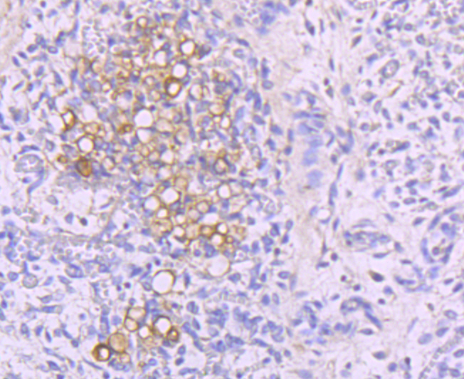
Immunohistochemical analysis of paraffin-embedded human thymus tissue using anti-NFAT2 antibody. Counter stained with hematoxylin.
Immunohistochemical analysis of paraffin-embedded human tonsil tissue using anti-NFAT2 antibody. Counter stained with hematoxylin.
Immunohistochemical analysis of paraffin-embedded human spleen tissue using anti-NFAT2 antibody. Counter stained with hematoxylin.
Immunohistochemical analysis of paraffin-embedded human skeleton muscle tissue using anti-NFAT2 antibody. Counter stained with hematoxylin.
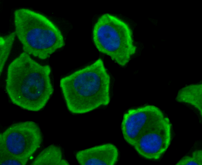
ICC staining NFAT2 in MCF-7 cells (green). The nuclear counter stain is DAPI (blue). Cells were fixed in paraformaldehyde, permeabilised with 0.25% Triton X100/PBS.
ICC staining NFAT2 in 293T cells (green). The nuclear counter stain is DAPI (blue). Cells were fixed in paraformaldehyde, permeabilised with 0.25% Triton X100/PBS.
ICC staining NFAT2 in Hela cells (green). The nuclear counter stain is DAPI (blue). Cells were fixed in paraformaldehyde, permeabilised with 0.25% Triton X100/PBS.
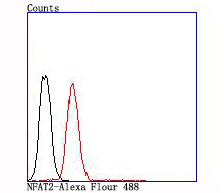
Flow cytometric analysis of Jurkat cells with NFAT2 antibody at 1/100 dilution (red) compared with an unlabelled control (cells without incubation with primary antibody; black).
Plays a role in the inducible expression of cytokine genes in T-cells, especially in the induction of the IL-2 or IL-4 gene transcription. Also controls gene expression in embryonic cardiac cells. Could regulate not only the activation and proliferation but also the differentiation and programmed death of T-lymphocytes as well as lymphoid and non-lymphoid cells. Required for osteoclastogenesis and regulates many genes important for osteoclast differentiation and function.
If you have published an article using product 49555, please notify us so that we can cite your literature.


 Yes
Yes


