Product Detail
Product NameDR5 Rabbit mAb
Clone No.JA03-38
Host SpeciesRecombinant Rabbit
Clonality Monoclonal
PurificationProA affinity purified
ApplicationsWB, IP, IHC, FC
Species ReactivityHu
Immunogen Descrecombinant protein
ConjugateUnconjugated
Other NamesFas like protein antibody
Apoptosis inducing protein TRICK2A/2B antibody
Apoptosis inducing receptor TRAIL R2 antibody
CD262 antibody
CD262 antigen antibody
Cytotoxic TRAIL receptor 2 antibody
Death domain containing receptor for TRAIL/Apo 2L antibody
Death receptor 5 antibody
DR5 antibody
KILLER antibody
KILLER/DR5 antibody
OTTHUMP00000123492 antibody
OTTHUMP00000123493 antibody
p53 regulated DNA damage inducible cell death receptor(killer) antibody
TNF related apoptosis inducing ligand receptor 2 antibody
TNF-related apoptosis-inducing ligand receptor 2 antibody
TNFRSF10B antibody
TR10B_HUMAN antibody
TRAIL R2 antibody
TRAIL receptor 2 antibody
TRAIL-R2 antibody
TRAILR2 antibody
TRICK2 antibody
TRICK2A antibody
TRICK2B antibody
TRICKB antibody
Tumor necrosis factor receptor like protein ZTNFR9 antibody
Tumor necrosis factor receptor superfamily member 10B antibody
Tumor necrosis factor receptor superfamily, member 10b antibody
ZTNFR9 antibody
Accession NoSwiss-Prot#:O14763
Uniprot
O14763
Gene ID
8795;
Calculated MW48 kDa
Formulation1*TBS (pH7.4), 1%BSA, 40%Glycerol. Preservative: 0.05% Sodium Azide.
StorageStore at -20˚C
Application Details
WB: 1:500-1:1,000
IHC: 1:50-1:200
FC: 1:50-1:100
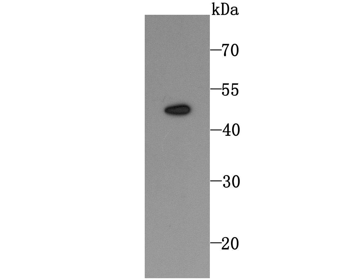
Western blot analysis of DR5 on HL-60 cell using anti-DR5 antibody at 1/1,000 dilution.
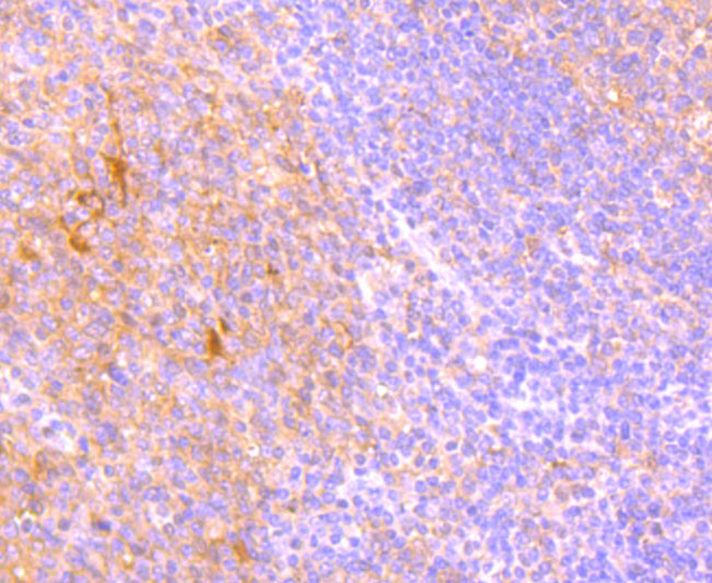
Immunohistochemical analysis of paraffin-embedded human tonsil tissue using anti-DR5 antibody. Counter stained with hematoxylin.
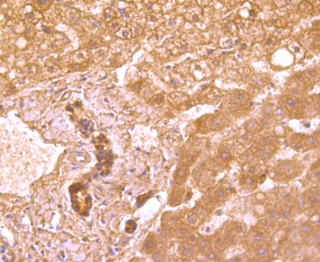
Immunohistochemical analysis of paraffin-embedded human liver tissue using anti-DR5 antibody. Counter stained with hematoxylin.
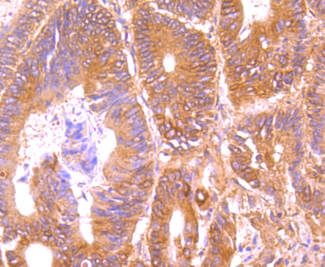
Immunohistochemical analysis of paraffin-embedded human colon cancer tissue using anti-DR5 antibody. Counter stained with hematoxylin.
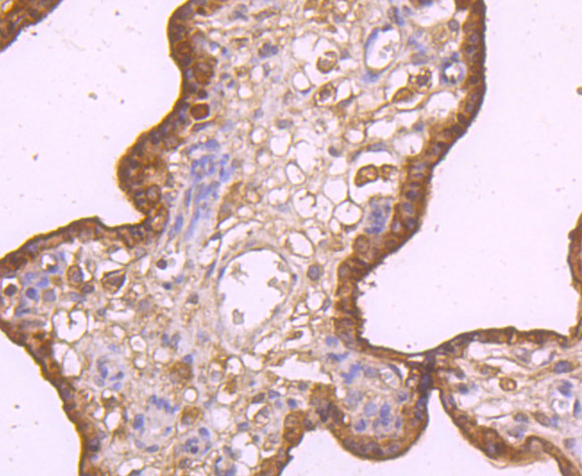
Immunohistochemical analysis of paraffin-embedded human placenta tissue using anti-DR5 antibody. Counter stained with hematoxylin.
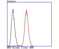
Flow cytometric analysis of SW480 cells with DR5 antibody at 1/100 dilution (red) compared with an unlabelled control (cells without incubation with primary antibody; black).
Tumor necrosis factor (TNF) is a pleiotropic cytokine whose function is mediated by two distinct cell surface receptors, designated TNF-R1 and TNF-R2, which are expressed on most cell types. TNF function is primarily mediated through TNF-R1 signaling. Both receptors belong to the growing TNF receptor superfamily which includes FAS antigen and CD40. TNF-R1 contains a cytoplasmic motif, termed the "death domain," that has been found to be necessary for the transduction of the apoptotic signal. The death domain is also found in several other receptors, including FAS, DR2 (or TRUNDD), DR3 (Death Receptor 3), DR4 and DR5. TRUNDD, DR4 and DR5 are receptors for the apoptosis-inducing cytokine TRAIL. A non-death domain-containing receptor, designated decoy receptor (DcR1 or TRID), also specifically associates with TRAIL and may play a role in cellular resistance to apoptotic stimuli.
If you have published an article using product 49563, please notify us so that we can cite your literature.


 Yes
Yes


