Product Detail
Product NameCLOCK Rabbit mAb
Clone No.JA12-33
Host SpeciesRecombinant Rabbit
Clonality Monoclonal
PurificationProA affinity purified
ApplicationsWB, ICC/IF, IHC, FC
Species ReactivityHu,Ms, Rt
Immunogen DescRecombinant protein
ConjugateUnconjugated
Other NamesbHLHe8 antibody
Circadian locomoter output cycles kaput protein antibody
Circadian locomoter output cycles protein kaput antibody
Circadian Locomotor Output Cycles Kaput antibody
Circadium Locomotor Output Cycles Kaput antibody
Class E basic helix-loop-helix protein 8 antibody
CLOCK antibody
Clock circadian regulator antibody
Clock homolog antibody
Clock protein antibody
CLOCK_HUMAN antibody
hCLOCK antibody
KIAA0334 antibody
Accession NoSwiss-Prot#:O15516
Uniprot
O15516
Gene ID
9575;
Formulation1*TBS (pH7.4), 1%BSA, 40%Glycerol. Preservative: 0.05% Sodium Azide.
StorageStore at -20˚C
Application Details
WB:1:500-1:1000
IHC: 1:50-1:200
ICC: 1:50-1:200
FC: 1:50-1:100
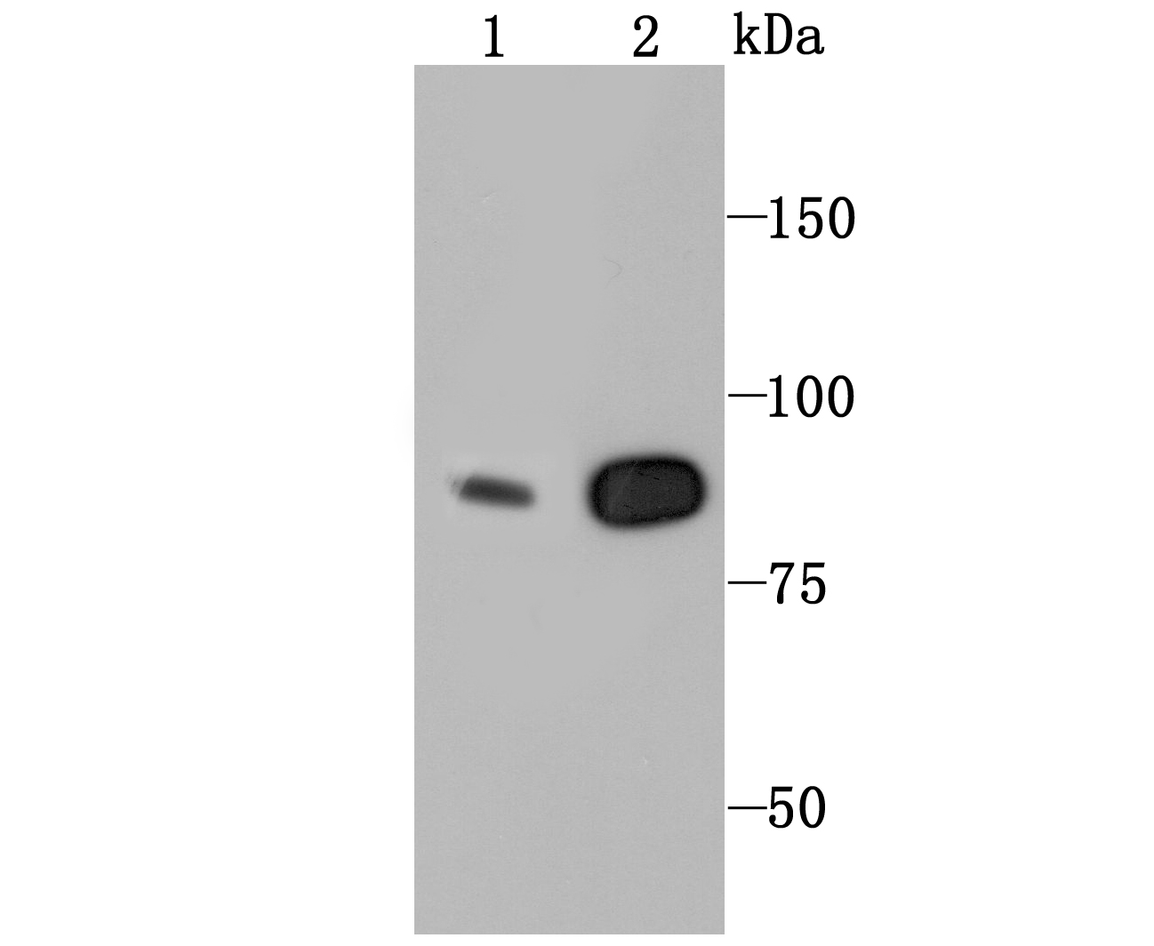
Western blot analysis of CLOCK on MCF-7 (1) and PC-12 (2) cell using anti-CLOCK antibody at 1/1,000 dilution.
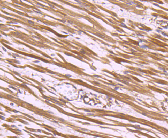
Immunohistochemical analysis of paraffin-embedded human fetal skeletal muscle tissue using anti-CLOCK antibody. Counter stained with hematoxylin.
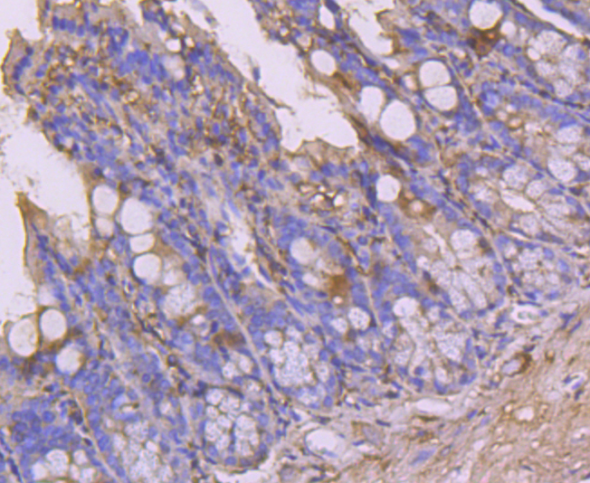
Immunohistochemical analysis of paraffin-embedded 2 colon tissue using anti-CLOCK antibody. Counter stained with hematoxylin.
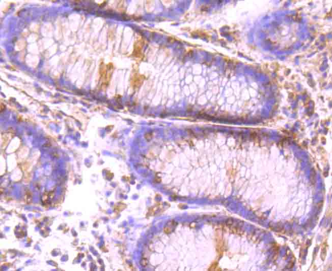
Immunohistochemical analysis of paraffin-embedded human colon cancer tissue using anti-CLOCK antibody. Counter stained with hematoxylin.
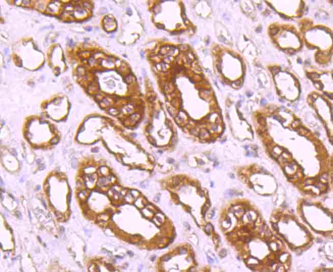
Immunohistochemical analysis of paraffin-embedded human kidney tissue using anti-CLOCK antibody. Counter stained with hematoxylin.
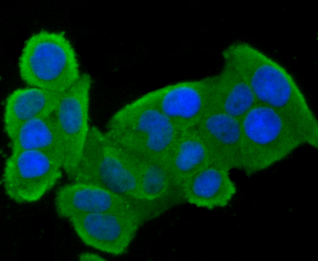
ICC staining CLOCK in Hela cells (green). The nuclear counter stain is DAPI (blue). Cells were fixed in paraformaldehyde, permeabilised with 0.25% Triton X100/PBS.
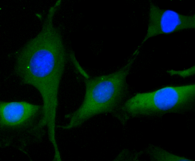
ICC staining CLOCK in SHG-44 cells (green). The nuclear counter stain is DAPI (blue). Cells were fixed in paraformaldehyde, permeabilised with 0.25% Triton X100/PBS.
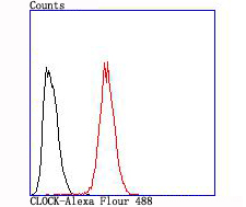
Flow cytometric analysis of Hela cells with CLOCK antibody at 1/100 dilution (red) compared with an unlabelled control (cells without incubation with primary antibody; black).
Biological timepieces called circadian clocks are responsible for the regulation of hormonal rhythms, sleep cycles and other behaviors. The superchiasmatic nucleus (SCN), which is located in the brain, was the first mammalian circadian clock to be discovered. Clock, a member of the Basic-helix-loop-helix-psp (bHLH-PAS) family of transcription factors, has also been identified as having circadian function. Mutations within the clock gene have been shown to increase the length of the endogenous period and To contain a loss of rhythmicity of circadian oscillations. Clock contains a DNA-binding domain, a protein dimerization domain and a glutamine-rich C-terminal region, which indicates transactivation ability. It has been speculated that Clock may regulation circadian rhythmicity in combination with Other proteins such as Per. Per is also a PAS-domain containing protein that exhibits circadian function. Highest expression of Clock is seen in the hypothalamus and the eye.
If you have published an article using product 49586, please notify us so that we can cite your literature.


 Yes
Yes


