Product Detail
Product NamePeroxiredoxin 3 Rabbit mAb
Clone No.JA53-21
Host SpeciesRecombinant Rabbit
Clonality Monoclonal
PurificationProA affinity purified
ApplicationsWB, ICC, IHC, IP, FC
Species ReactivityHu
Immunogen Descrecombinant protein
ConjugateUnconjugated
Other NamesAntioxidant protein 1 antibody
AOP 1 antibody
AOP-1 antibody
AOP1 antibody
HBC189 antibody
MER5 antibody
MGC104387 antibody
MGC24293 antibody
mitochondrial antibody
peroxiredoxin 3 antibody
Peroxiredoxin III antibody
Peroxiredoxin-3 antibody
PRDX3 antibody
PRDX3_HUMAN antibody
PRO1748 antibody
Protein MER5 homolog antibody
PRX III antibody
Prx-III antibody
PRX3 antibody
SP 22 antibody
SP-22 antibody
SP22 antibody
Thioredoxin dependent peroxide reductase mitochondrial antibody
Thioredoxin-dependent peroxide reductase antibody
Accession NoSwiss-Prot#:P30048
Uniprot
P30048
Gene ID
10935;
Calculated MW24 kDa
Formulation1*TBS (pH7.4), 1%BSA, 40%Glycerol. Preservative: 0.05% Sodium Azide.
StorageStore at -20˚C
Application Details
WB: 1:500-1:2,000
IHC: 1:50-1:200
ICC: 1:50-1:200
FC: 1:50-1:100
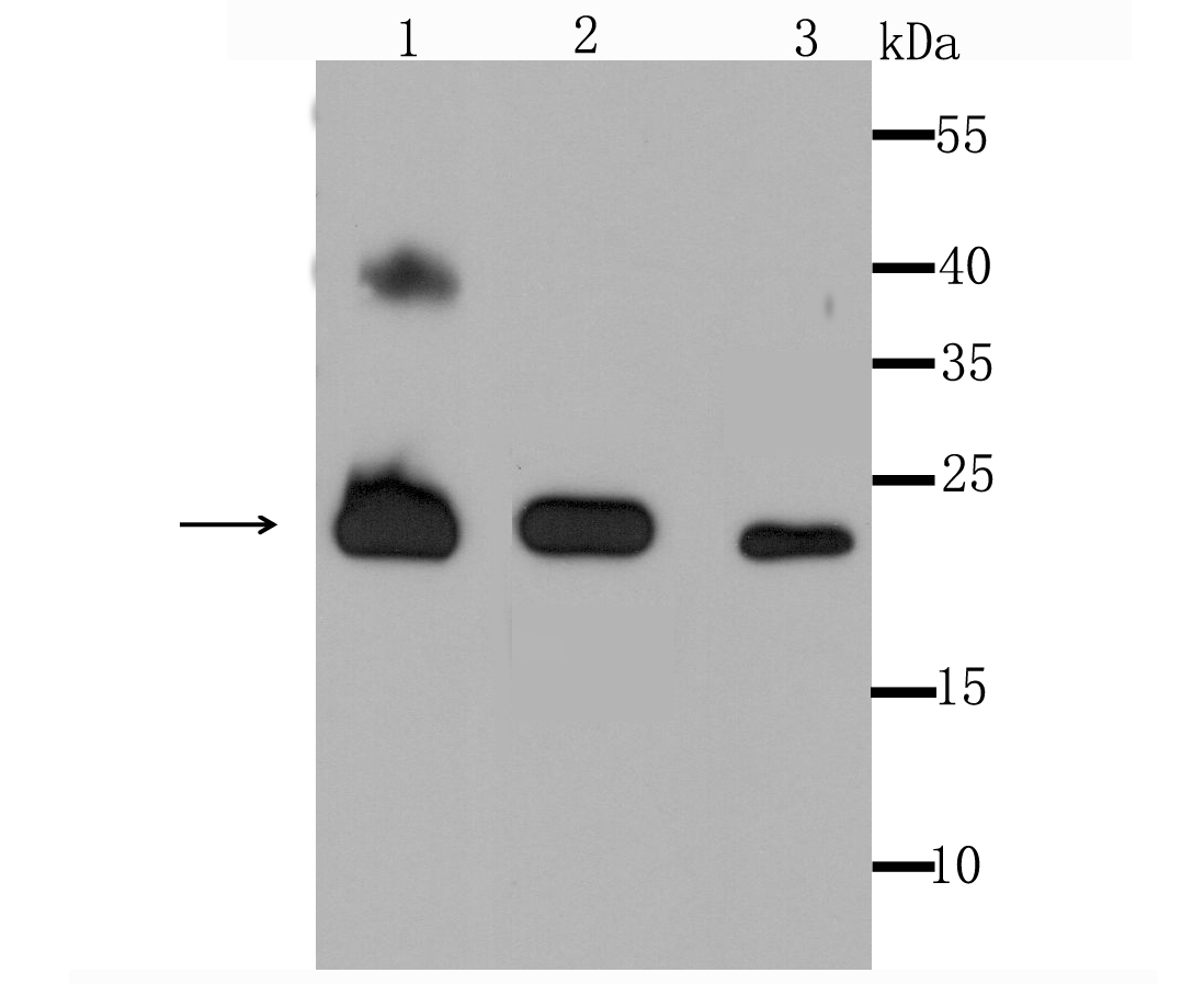
Western blot analysis of Peroxiredoxin 3 on different cell lysate using anti-Peroxiredoxin 3 antibody at 1/1,000 dilution. Positive control��
Lane1: Human liver
Lane2: MCF-7
Lane3: A431
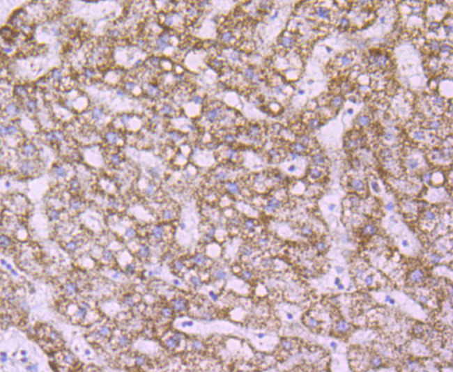
Immunohistochemical analysis of paraffin-embedded human liver tissue using anti- Peroxiredoxin 3 antibody. Counter stained with hematoxylin.
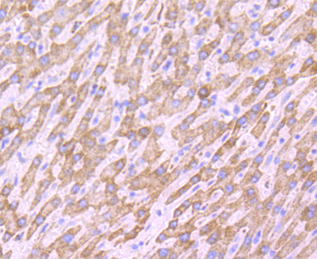
Immunohistochemical analysis of paraffin-embedded human liver cancer tissue using anti- Peroxiredoxin 3 antibody. Counter stained with hematoxylin.
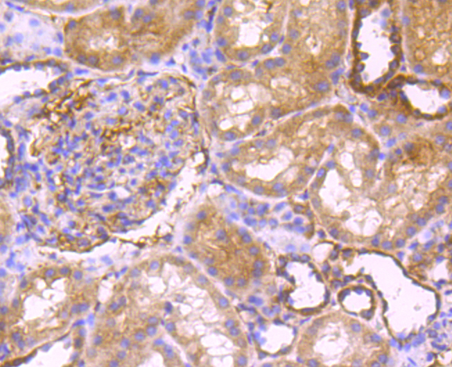
Immunohistochemical analysis of paraffin-embedded human kidney tissue using anti- Peroxiredoxin 3 antibody. Counter stained with hematoxylin.
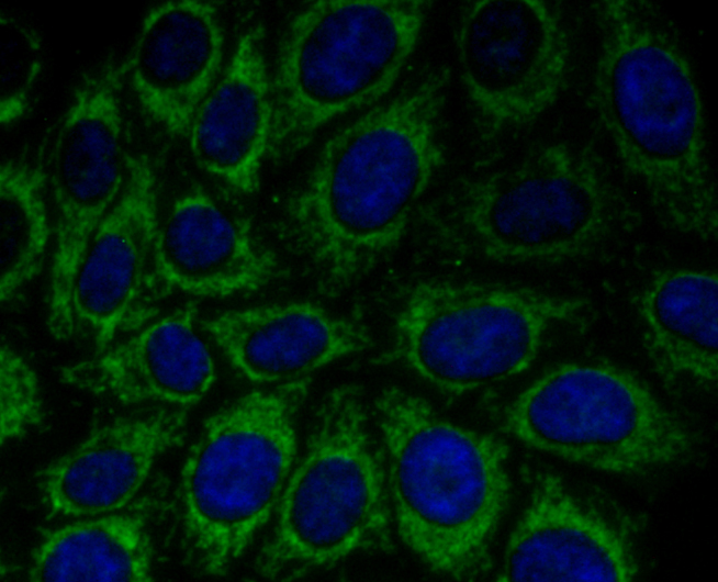
ICC staining Peroxiredoxin 3 in Hela cells (green). The nuclear counter stain is DAPI (blue). Cells were fixed in paraformaldehyde, permeabilised with 0.25% Triton X100/PBS.
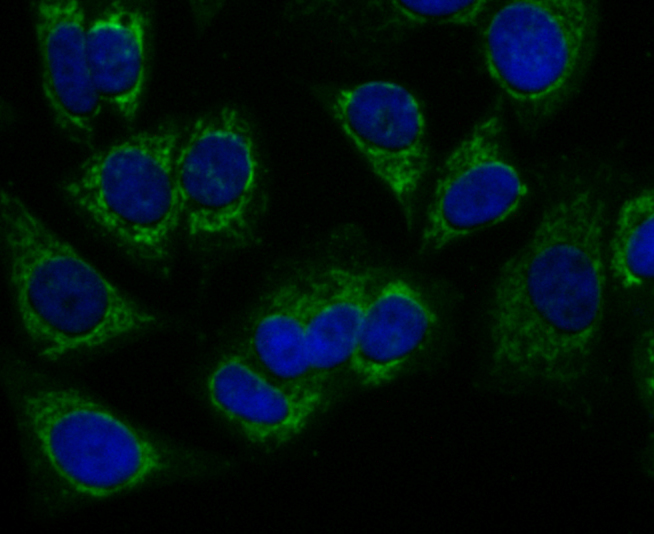
ICC staining Peroxiredoxin 3 in HepG2 cells (green). The nuclear counter stain is DAPI (blue). Cells were fixed in paraformaldehyde, permeabilised with 0.25% Triton X100/PBS.
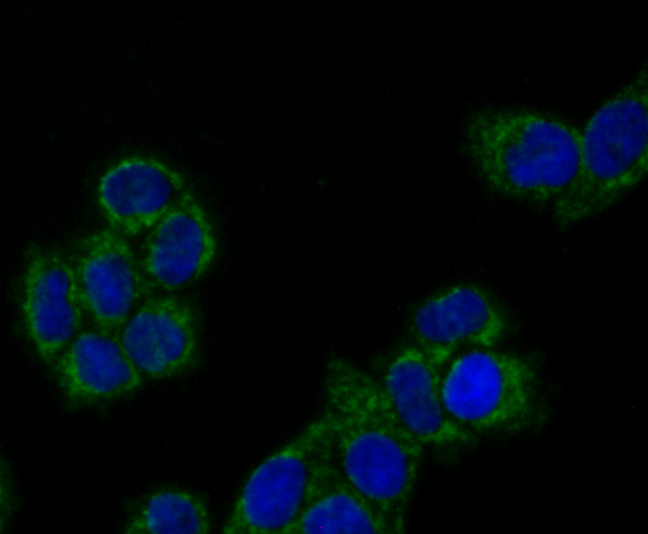
ICC staining Peroxiredoxin 3 in MCF-7 cells (green). The nuclear counter stain is DAPI (blue). Cells were fixed in paraformaldehyde, permeabilised with 0.25% Triton X100/PBS.
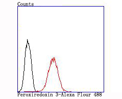
Flow cytometric analysis of MCF-7 cells with Peroxiredoxin 3 antibody at 1/100 dilution (red) compared with an unlabelled control (cells without incubation with primary antibody; black).
The peroxiredoxin (PRX) family comprises six antioxidant proteins, PRX I, II, III, IV, V and VI, which protect cells from reactive oxygen species (ROS) by preventing the metal-catalyzed oxidation of enzymes. The PRX proteins primarily utilize thioredoxin as the electron donor for antioxidation, although they are fairly promiscuous with regard to the hydroperoxide substrate. In addition to protection from ROS, peroxiredoxins are also involved in cell proliferation, differentiation and gene expression. PRX I, II, IV and VI show diffuse cytoplasmic localization, while PRX III and V exhibit distinct mitochondrial localization. The human PRX I gene encodes a protein that is expressed in several tissues, including liver, kidney, testis, lung and nervous system. PRX II is expressed in testis, while PRX III shows expression in lung. PRX I, II and III are overexpressed in breast cancer and may be involved in its development or progression. Upregulated protein levels of PRX I and II in Alzheimer's disease (AD) and Down syndrome (DS) indicate the involvement of PRX I and II in their pathogenesis. The human PRX IV gene is abundantly expressed in many tissues.
If you have published an article using product 49595, please notify us so that we can cite your literature.


 Yes
Yes


