Location:
Home
>
Recombinant Rabbit Monoclonal Antibodies > Pyruvate Dehydrogenase E1 beta subunit Rabbit mAb
Pyruvate Dehydrogenase E1 beta subunit Rabbit mAb#49646

 Yes
Yes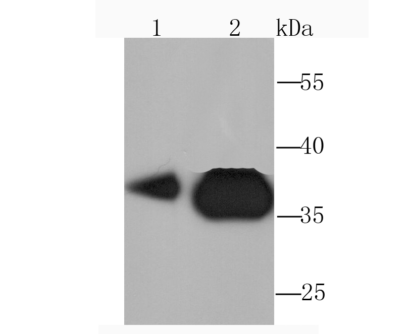
Western blot analysis of Pyruvate Dehydrogenase E1 beta subunit on HepG2 (1) and Hela (2) cell lysate using anti-Pyruvate Dehydrogenase E1 beta subunit antibody at 1/1,000 dilution.
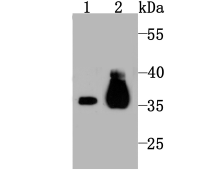
Western blot analysis of Pyruvate Dehydrogenase E1 beta subunit on mouse kidney tissue (1) and PC-12 (2) cell lysate using anti-Pyruvate Dehydrogenase E1 beta subunit antibody at 1/1,000 dilution.
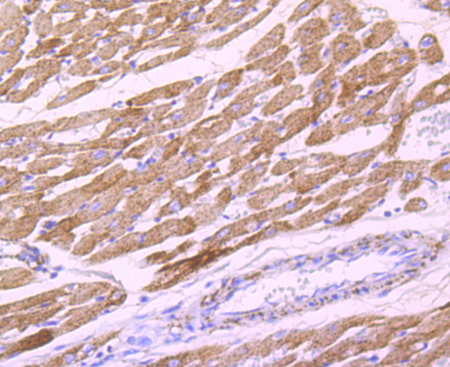
Immunohistochemical analysis of paraffin-embedded rat heart tissue using anti-Pyruvate Dehydrogenase E1 beta subunit antibody. Counter stained with hematoxylin.
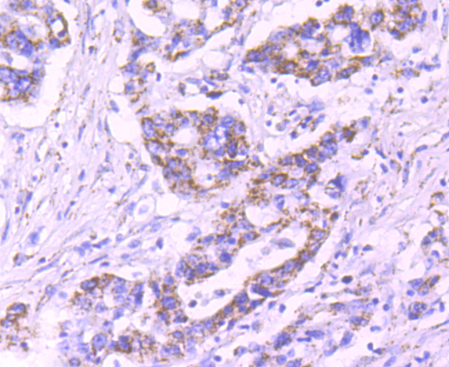
Immunohistochemical analysis of paraffin-embedded human stomach cancer tissue using anti-Pyruvate Dehydrogenase E1 beta subunit antibody. Counter stained with hematoxylin.
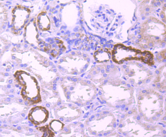
Immunohistochemical analysis of paraffin-embedded human kidney tissue using anti-Pyruvate Dehydrogenase E1 beta subunit antibody. Counter stained with hematoxylin.
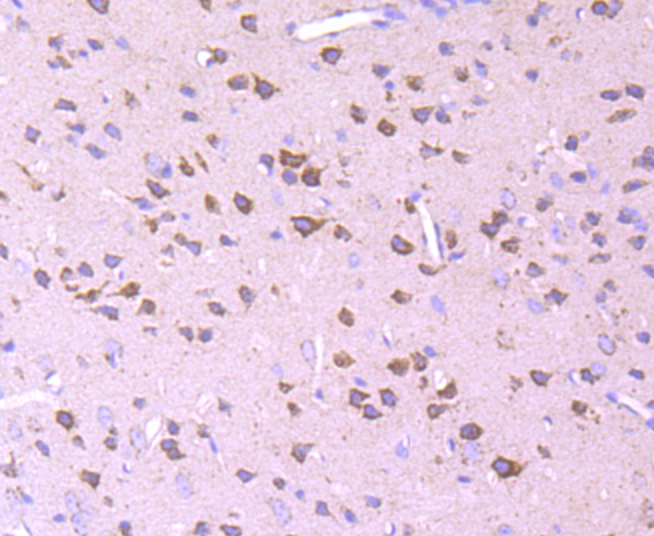
Immunohistochemical analysis of paraffin-embedded mouse brain tissue using anti-Pyruvate Dehydrogenase E1 beta subunit antibody. Counter stained with hematoxylin.
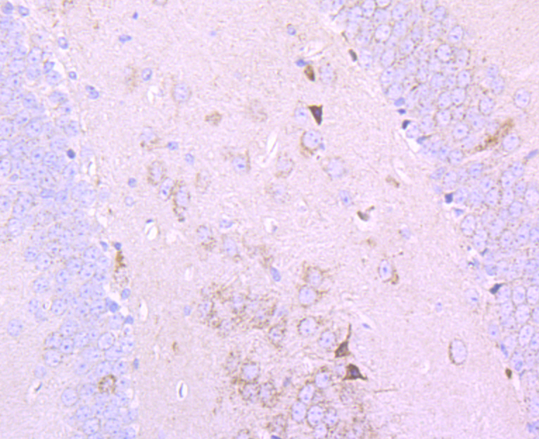
Immunohistochemical analysis of paraffin-embedded mouse hippocampus tissue using anti-Pyruvate Dehydrogenase E1 beta subunit antibody. Counter stained with hematoxylin.
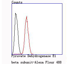
Flow cytometric analysis of Hela cells with Pyruvate Dehydrogenase E1 beta subunit antibody at 1/100 dilution (red) compared with an unlabelled control (cells without incubation with primary antibody; black).
NOTE
Application
- WBWestern Blotting
- IHCImmunohistochemistry
- IFImmunofluorescence
- ICCImmunocytochemistry
- FCFlow Cytometry
- IPImmunoprecipitation
- EELISA
- DBDot Blotting
- ChIPChromatin Immunoprecipitation
- GICAGold Immunochromatography Assay
- NCNegative Control
Species Reactivity
- HuHuman
- MsMouse
- RtRat
- DmDrosophila melanogaster
- CCaenorhabditis elegans
- MkMonkey
- RbRabbit
- BBovine
- DDog
- PPig
- HmHamster
- ChHmChinese Hamster
- ChkChicken
- ShpSheep


