Welcome to Sabbiotech!     
Email:info@signalwayantibody.com
Location: Home > Recombinant Rabbit Monoclonal Antibodies > Iba1 Rabbit mAb

Iba1 Rabbit mAb#49668

Availability: Yes
Total Amount:$
Product Detail

Product NameIba1 Rabbit mAb

Clone No.JM36-62

ClonalityMonoclonal

PurificationProA affinity purified

ApplicationsWB, ICC,IF, IHC, FC, IP

Species ReactivityHu, Ms, Rt

Immunogen DescRecombinant protein

ConjugateUnconjugated

Other NamesAIF 1 antibody
AIF-1 antibody
Aif1 antibody
AIF1 protein antibody
AIF1_HUMAN antibody
Allograft inflammatory factor 1 antibody
Allograft inflammatory factor 1 splice variant G antibody
allograft inflammatory factor-1 splice variant Hara-1 antibody
balloon angioplasty responsive transcription antibody
BART 1 antibody
G1 antibody
G1 putative splice variant of allograft inflamatory factor 1 antibody
IBA 1 antibody
IBA1 antibody
interferon gamma responsive transcript antibody
Interferon responsive transcript 1 antibody
interferon responsive transcript factor 1 antibody
Ionized calcium binding adapter molecule 1 antibody
Ionized calcium-binding adapter molecule 1 antibody
ionized calcium-binding adapter molecule antibody
IRT 1 antibody
IRT1 antibody
Microglia response factor antibody
MRF1 antibody
Protein g1 antibody

Accession NoSwiss-Prot#:P55008

Uniprot P55008

Gene ID 199;

Calculated MW17 kDa

Formulation1*TBS (pH7.4), 1%BSA, 40%Glycerol. Preservative: 0.05% Sodium Azide.

StorageStore at -20˚C

Application Details

WB: 1:500-1:1,000
IHC: 1:100-1:500
ICC/IF: 1:50-1:100
IP: 1:10-1:50
FC: 1:50-1:200

Immunohistochemical analysis of paraffin-embedded rat lung tissue using anti-Iba1 antibody. Counter stained with hematoxylin.
Immunohistochemical analysis of paraffin-embedded human lung cancer tissue using anti-Iba1 antibody. Counter stained with hematoxylin.
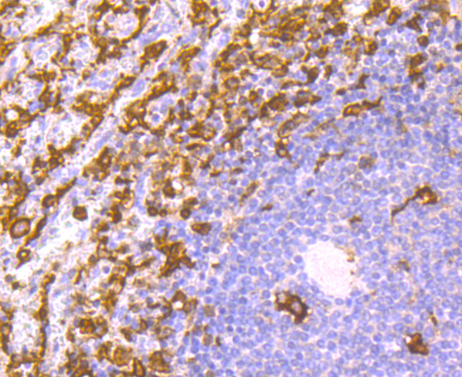
Immunohistochemical analysis of paraffin-embedded human spleen tissue using anti-Iba1 antibody. Counter stained with hematoxylin.
Immunohistochemical analysis of paraffin-embedded mouse brain tissue using anti-Iba1 antibody. Counter stained with hematoxylin.
Immunohistochemical analysis of paraffin-embedded mouse spleen tissue using anti-Iba1 antibody. Counter stained with hematoxylin.
ICC staining Iba1 in SH-SY5Y cells (green). The nuclear counter stain is DAPI (blue). Cells were fixed in paraformaldehyde, permeabilised with 0.25% Triton X100/PBS.
Flow cytometric analysis of THP-1 cells with Iba1 antibody at 1/100 dilution (red) compared with an unlabelled control (cells without incubation with primary antibody; black).

Ionized calcium-binding adapter molecule 1 (Iba1), also known as allograft inflammatory factor-1 (AIF-1), is a 147 amino acid cytoplasmic, calcium-binding protein that is thought to play a role in macrophage activation and function. Iba1, containing two EF domains, is induced by cytokines and interferons. In an unstimulated state, Iba1 colocalizes with actin, and upon stimulation, translocates to lamellipodia. It is also a marker of human microglia and is expressed by macrophages in injured skeletal muscle. The gene encoding Iba1 maps to chromosome 6p21.33 and resides in the tumor necrosis factor (TNF) cluster of genes located in the region represented by the human major histocompatibility complex (MHC).

If you have published an article using product 49668, please notify us so that we can cite your literature.

NOTE

Application

  • WBWestern Blotting
  • IHCImmunohistochemistry
  • IFImmunofluorescence
  • ICCImmunocytochemistry
  • FCFlow Cytometry
  • IPImmunoprecipitation
  • EELISA
  • DBDot Blotting
  • ChIPChromatin Immunoprecipitation
  • GICAGold Immunochromatography Assay
  • NCNegative Control

Species Reactivity

  • HuHuman
  • MsMouse
  • RtRat
  • DmDrosophila melanogaster
  • CCaenorhabditis elegans
  • MkMonkey
  • RbRabbit
  • BBovine
  • DDog
  • PPig
  • HmHamster
  • ChHmChinese Hamster
  • ChkChicken
  • ShpSheep
;