Welcome to Sabbiotech!     
Email:info@signalwayantibody.com
Location: Home > Recombinant Rabbit Monoclonal Antibodies > NRF1 Rabbit mAb

NRF1 Rabbit mAb#49676

Availability: Yes
Total Amount:$
Product Detail

Product NameNRF1 Rabbit mAb

Clone No.JM89-63

Host SpeciesRecombinant Rabbit

Clonality Monoclonal

PurificationProA affinity purified

ApplicationsWB, ICC/IF, IHC, FC, IP

Species ReactivityHu, Ms, Rt

Immunogen DescRecombinant protein

ConjugateUnconjugated

Other Namesalpha pal antibody
alpha palindromic binding protein antibody
Alpha palindromic-binding protein antibody
Alpha-pal antibody
locus control region factor 1 antibody
NFE2 related factor 1 antibody
NRF-1 antibody
Nrf1 antibody
NRF1_HUMAN antibody
Nuclear respiratory factor 1 antibody

Accession NoSwiss-Prot#:Q16656

Uniprot Q16656

Gene ID 4899;

Calculated MW56 kDa/65kDa(Observed)

Formulation1*TBS (pH7.4), 1%BSA, 40%Glycerol. Preservative: 0.05% Sodium Azide.

StorageStore at -20˚C

Application Details
WB: 1:500
IHC: 1:50-1:200
ICC: 1:50-1:200

FC: 1:50-1:100
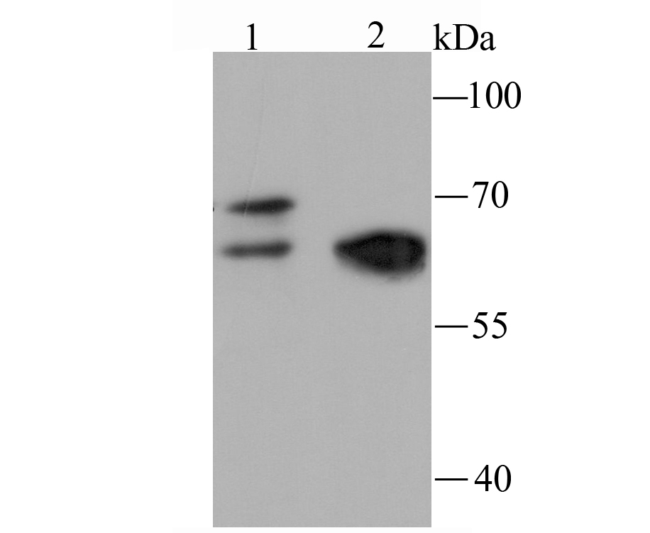
Western blot analysis of NRF1 on Hela cell (1) and mouse heart tissue (2) lysate using anti-NRF1 antibody at 1/500 dilution.
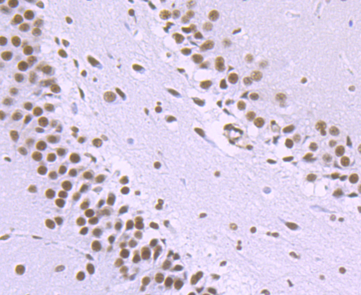
Immunohistochemical analysis of paraffin-embedded rat brain tissue using anti-NRF1 antibody. Counter stained with hematoxylin.
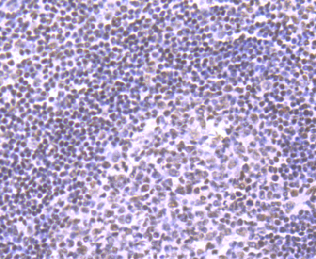
Immunohistochemical analysis of paraffin-embedded human tonsil tissue using anti-NRF1 antibody. Counter stained with hematoxylin.
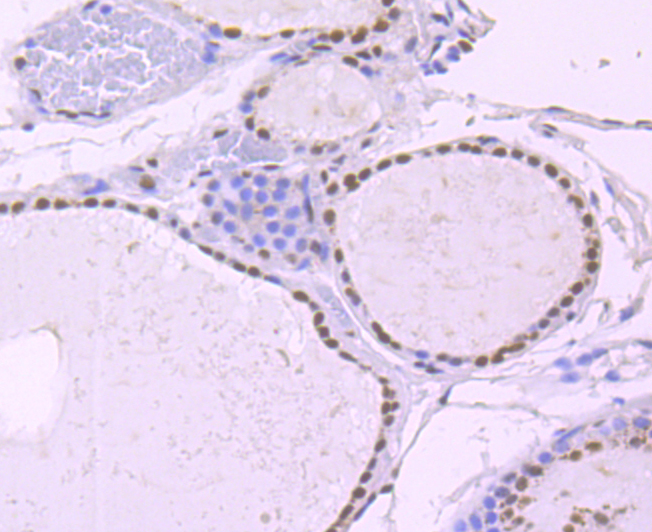
Immunohistochemical analysis of paraffin-embedded human thyriod tissue using anti-NRF1 antibody. Counter stained with hematoxylin.
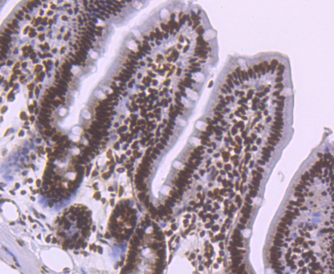
Immunohistochemical analysis of paraffin-embedded mouse colon tissue using anti-NRF1 antibody. Counter stained with hematoxylin.
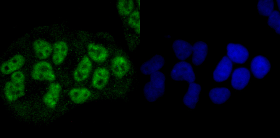
ICC staining NRF1 in Hela cells (green). The nuclear counter stain is DAPI (blue). Cells were fixed in paraformaldehyde, permeabilised with 0.25% Triton X100/PBS.
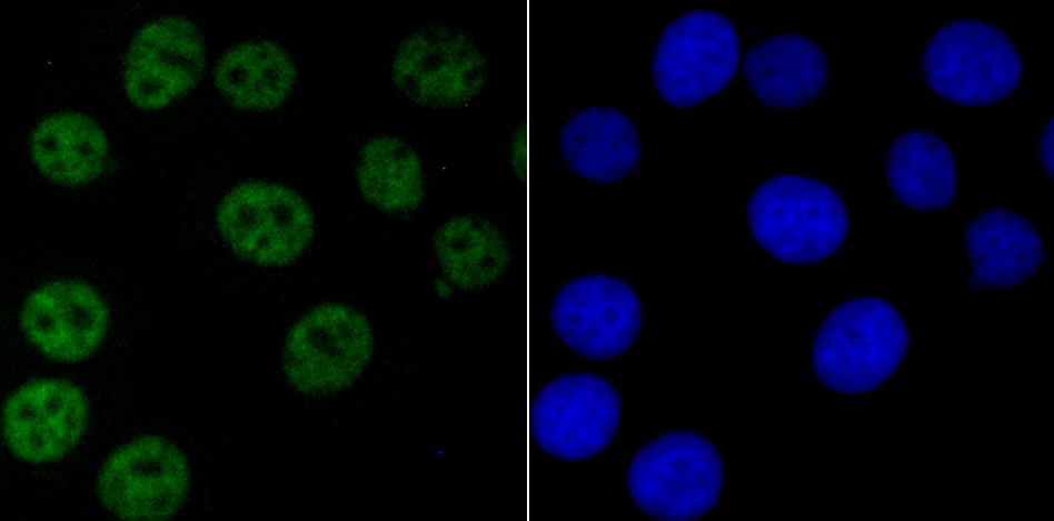
ICC staining NRF1 in MCF-7 cells (green). The nuclear counter stain is DAPI (blue). Cells were fixed in paraformaldehyde, permeabilised with 0.25% Triton X100/PBS.
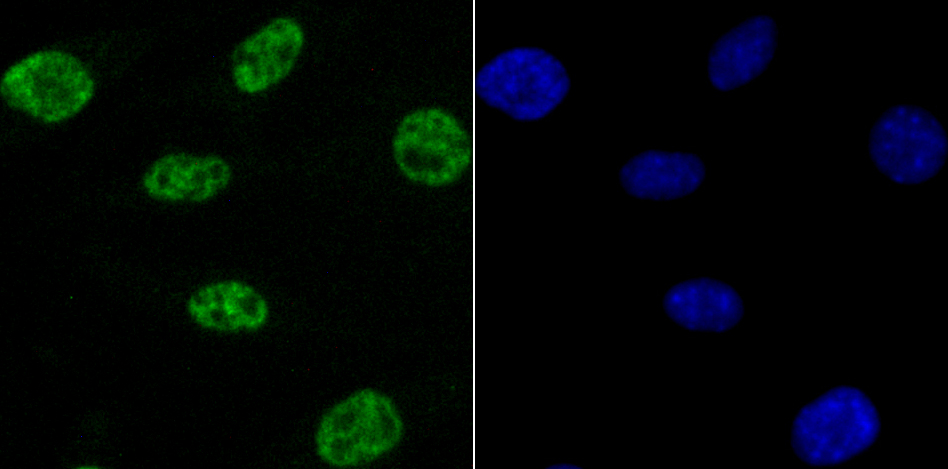
ICC staining NRF1 in SH-SY5Y cells (green). The nuclear counter stain is DAPI (blue). Cells were fixed in paraformaldehyde, permeabilised with 0.25% Triton X100/PBS.
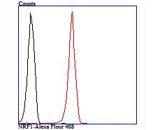
Flow cytometric analysis of 293T cells with NRF1 antibody at 1/100 dilution (red) compared with an unlabelled control (cells without incubation with primary antibody; black).
The NF-E2 DNA binding protein is composed of two subunits, p45 and MafK, and it regulates expression of globin genes in developing erythroid cells through interaction with Maf recognition elements (MAREs). A family of NF-E2 related proteins, which are collectively known as the Cap 'n' collar (CNC) family and include Nrf1 (also designated TCF11), Nrf2 and Nrf3, are bZIP transcription factors that heterodimerize with Maf proteins to bind MARE sequences. The Nrf proteins also bind the antioxidant response element (ARE) and are implicated in the regulation of detoxification enzymes and the oxidative stress response. They do so by heterodimerizing with Jun family members (c-Jun, JunB and JunD) to activate gene expression, specifically the detoxifying enzyme, NQO1. Nrf2 is widely expressed and is thought to translocate to the nucleus after treatment with xenobiotics and antioxidants, which stimulate its release from a repressor protein Keap1. Nrf3 is highly expressed in placenta, B cells and monocytes.

If you have published an article using product 49676, please notify us so that we can cite your literature.

NOTE

Application

  • WBWestern Blotting
  • IHCImmunohistochemistry
  • IFImmunofluorescence
  • ICCImmunocytochemistry
  • FCFlow Cytometry
  • IPImmunoprecipitation
  • EELISA
  • DBDot Blotting
  • ChIPChromatin Immunoprecipitation
  • GICAGold Immunochromatography Assay
  • NCNegative Control

Species Reactivity

  • HuHuman
  • MsMouse
  • RtRat
  • DmDrosophila melanogaster
  • CCaenorhabditis elegans
  • MkMonkey
  • RbRabbit
  • BBovine
  • DDog
  • PPig
  • HmHamster
  • ChHmChinese Hamster
  • ChkChicken
  • ShpSheep
;