Product Detail
Product NameTLR2 Rabbit mAb
Clone No.SR4549
Host SpeciesRecombinant Rabbit
ClonalityMonoclonal
PurificationProA affinity purified
ApplicationsWB, ICC/IF, IHC, FC
Species ReactivityHu, Ms, Rt
Immunogen DescRecombinant protein
ConjugateUnconjugated
Other NamesCD282 antibody
CD282 antigen antibody
TIL 4 antibody
TIL4 antibody
TLR 2 antibody
TLR2 antibody
TLR2_HUMAN antibody
Toll like receptor 2 antibody
Toll like receptor 2 precursor antibody
Toll-like receptor 2 antibody
Toll/interleukin 1 receptor like 4 antibody
Toll/interleukin 1 receptor like protein 4 antibody
Toll/interleukin receptor like protein 4 antibody
Toll/interleukin-1 receptor-like protein 4 antibody
Accession NoSwiss-Prot#:O60603
Uniprot
O60603
Gene ID
7097;
Formulation1*TBS (pH7.4), 1%BSA, 40%Glycerol. Preservative: 0.05% Sodium Azide.
StorageStore at -20˚C
Application Details
IHC: 1:50-1:200
ICC: 1:50-1:200
FC: 1:50-1:100
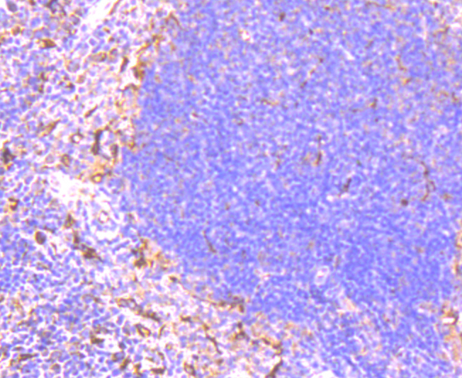
Immunohistochemical analysis of paraffin-embedded mouse spleen tissue using anti-TLR2 antibody. Counter stained with hematoxylin.
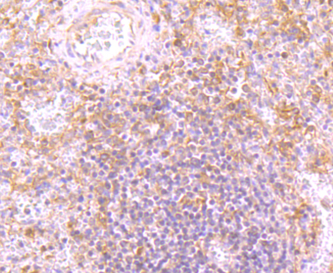
Immunohistochemical analysis of paraffin-embedded human spleen tissue using anti-TLR2 antibody. Counter stained with hematoxylin.
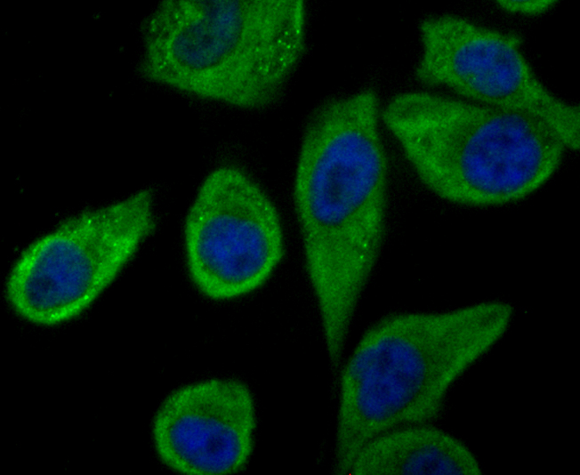
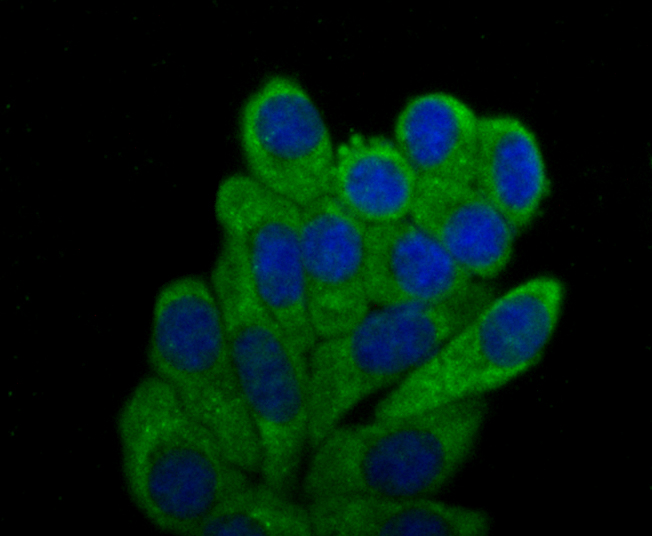
ICC staining TLR2 in PC-3M cells (green). The nuclear counter stain is DAPI (blue). Cells were fixed in paraformaldehyde, permeabilised with 0.25% Triton X100/PBS.
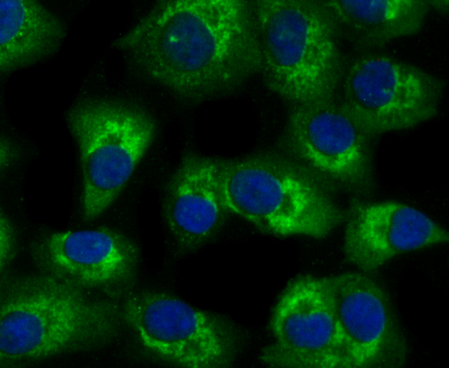
ICC staining TLR2 in A549 cells (green). The nuclear counter stain is DAPI (blue). Cells were fixed in paraformaldehyde, permeabilised with 0.25% Triton X100/PBS.
ICC staining TLR2 in HepG2 cells (green). The nuclear counter stain is DAPI (blue). Cells were fixed in paraformaldehyde, permeabilised with 0.25% Triton X100/PBS.
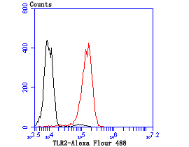
Flow cytometric analysis of THP-1 cells with TLR2 antibody at 1/100 dilution (red) compared with an unlabelled control (cells without incubation with primary antibody; black).
The TLR family of proteins are characterized by a highly conserved Toll homology (TH) domain, which is essential for Toll-induced signal transduction. TLR1, as well as the other TLR family members, are type I transmembrane receptors that characteristically contain an extracellular domain consisting of several leucine-rich regions along with a single cytoplasmic Toll/IL-1R-like domain. TLR2 and TLR4 are activated in response to lipopolysacchride (LPS) stimulation, which results in the activation and translocation of NFkB and suggests that these receptors are involved in mediating inflammatory responses. Expression of TLR receptors is highest in peripheral blood leukocytes, macrophages, and monocytes. TLR6 is highly homologous to TLR1, sharing greater than 65% sequence identity, and, like other members of TLR family, it induces NFkB signaling upon activation.
If you have published an article using product 49683, please notify us so that we can cite your literature.


 Yes
Yes


