Location:
Home
>
Recombinant Rabbit Monoclonal Antibodies > Vinculin Rabbit mAb
Vinculin Rabbit mAb#49684

 Yes
Yes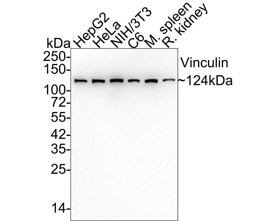
Western blot analysis of Vinculin on different lysates with Vinculin antibody at 1/20,000 dilution.
Lane 1: HepG2 cell lysate (15 µg/Lane)
Lane 2: HeLa cell lysate (15 µg/Lane)
Lane 3: NIH/3T3 cell lysate (15 µg/Lane)
Lane 4: C6 cell lysate (15 µg/Lane)
Lane 5: Mouse spleen tissue lysate (20 µg/Lane)
Lane 6: Rat kidney tissue lysate (20 µg/Lane)
Predicted band size: 124 kDa
Observed band size: 124 kDa
Exposure time: 1 minute 21 seconds;
4-20% SDS-PAGE gel.
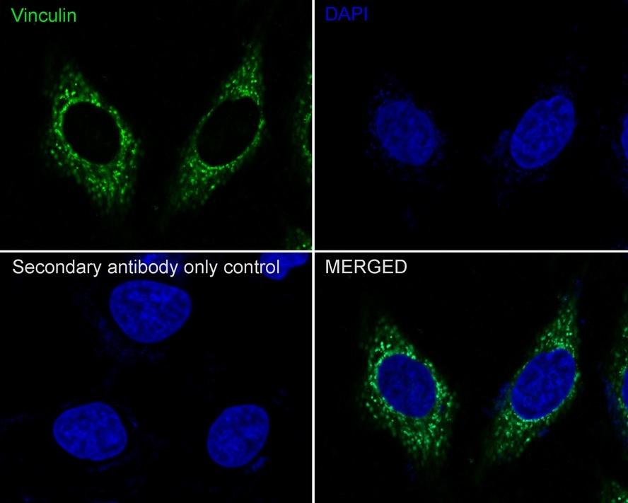
Immunocytochemistry analysis of HeLa cells labeling Vinculin with Vinculin antibody at 1/1,000 dilution.
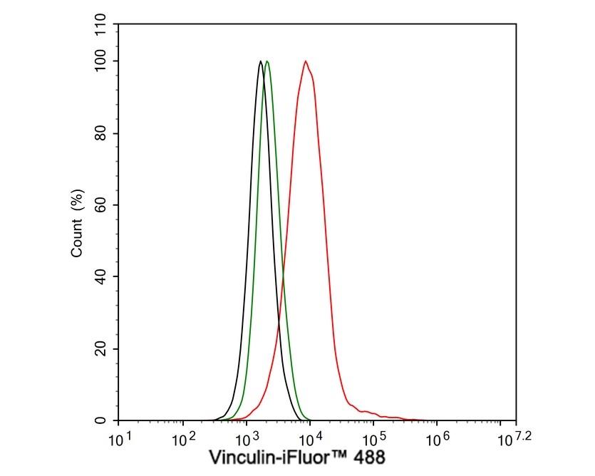
Flow cytometric analysis of HeLa cells labeling Vinculin.

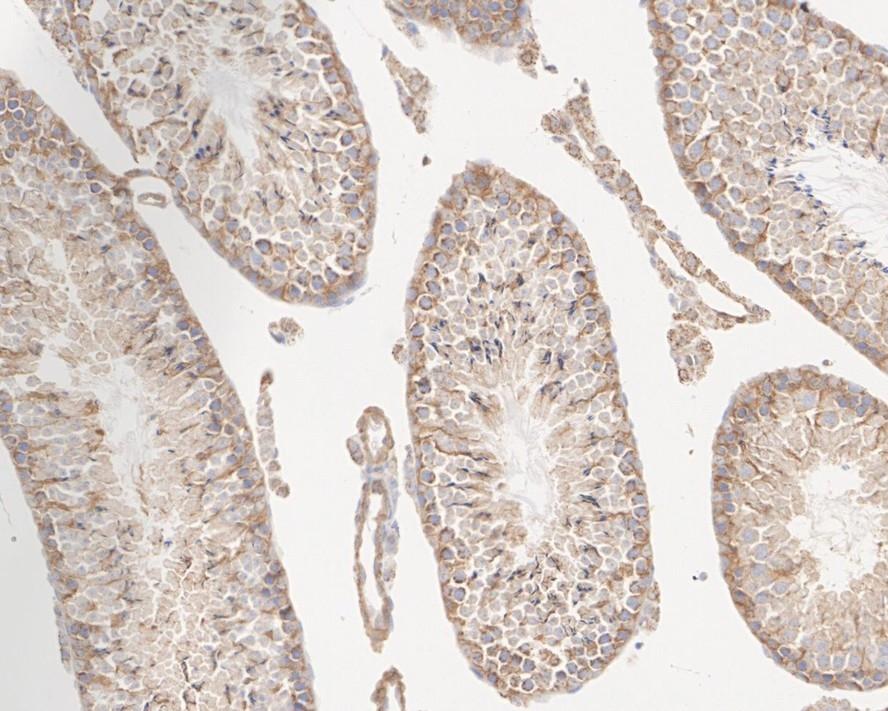
Immunohistochemical analysis of paraffin-embedded human testis tissue with Vinculin antibody at 1/200 dilution.
Immunohistochemical analysis of paraffin-embedded mouse testis tissue with Vinculin antibody at 1/20,000 dilution.
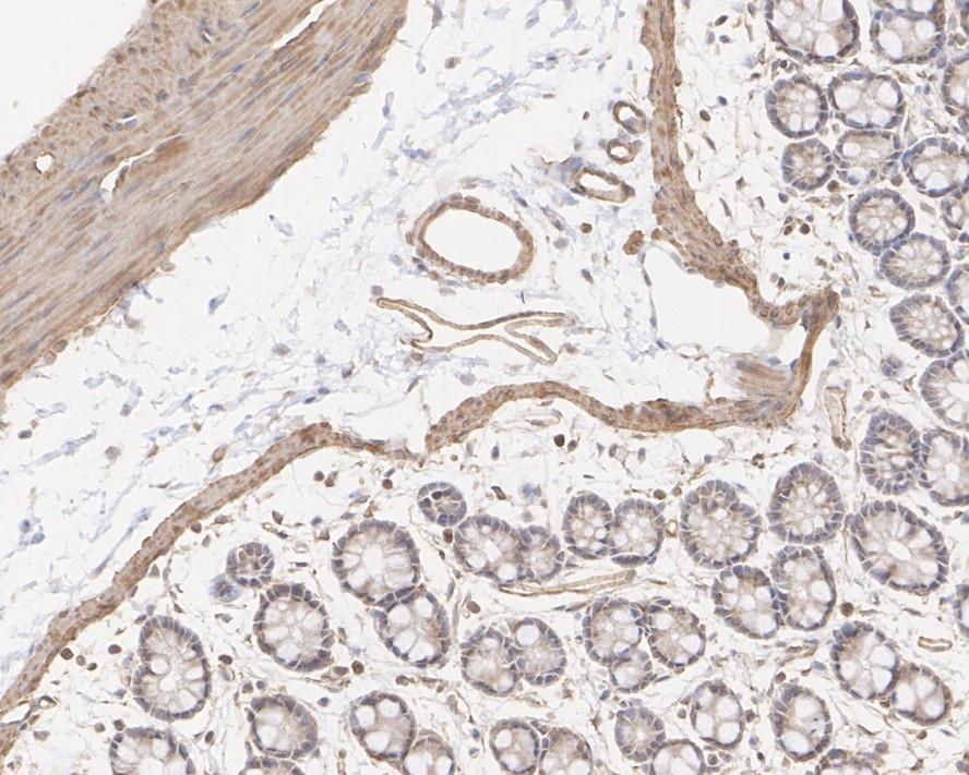
Immunohistochemical analysis of paraffin-embedded rat colon tissue with Vinculin antibody at 1/20,000 dilution.
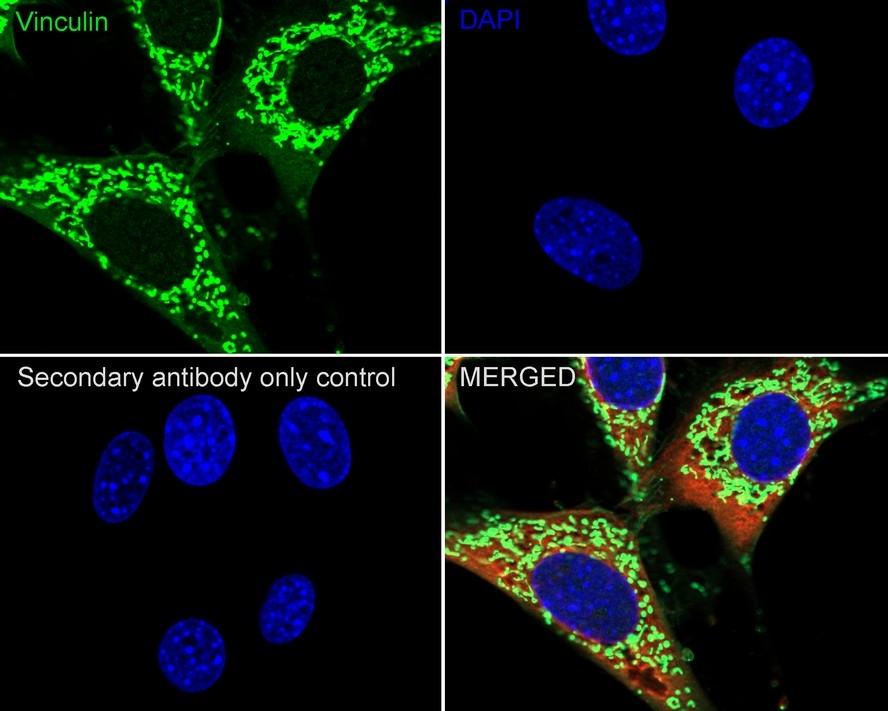
Immunocytochemistry analysis of NIH/3T3 cells labeling Vinculin with Vinculin antibody at 1/250 dilution.
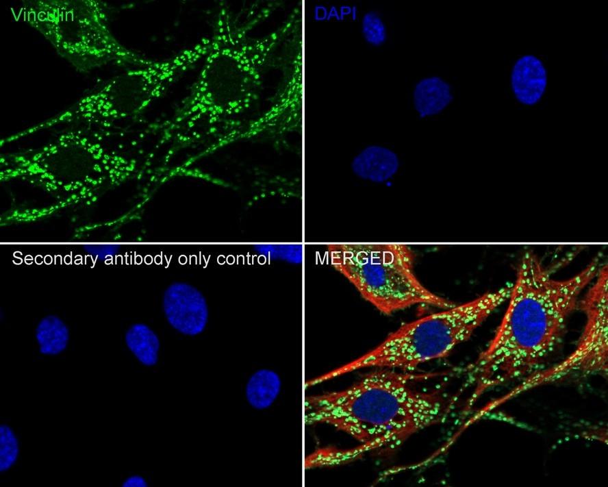
Immunocytochemistry analysis of C6 cells labeling Vinculin with Vinculin antibody at 1/250 dilution.
NOTE
Application
- WBWestern Blotting
- IHCImmunohistochemistry
- IFImmunofluorescence
- ICCImmunocytochemistry
- FCFlow Cytometry
- IPImmunoprecipitation
- EELISA
- DBDot Blotting
- ChIPChromatin Immunoprecipitation
- GICAGold Immunochromatography Assay
- NCNegative Control
Species Reactivity
- HuHuman
- MsMouse
- RtRat
- DmDrosophila melanogaster
- CCaenorhabditis elegans
- MkMonkey
- RbRabbit
- BBovine
- DDog
- PPig
- HmHamster
- ChHmChinese Hamster
- ChkChicken
- ShpSheep


