Product Detail
Product NameSATB2 Rabbit mAb
Host SpeciesRecombinant Rabbit
Clonality Monoclonal
PurificationProA affinity purified
ApplicationsWB, ICC/IF, IHC, FC
Species ReactivityHu, Ms, Rt
Immunogen DescRecombinant protein
ConjugateUnconjugated
Other NamesDNA binding protein SATB2 antibody
DNA-binding protein SATB2 antibody
FLJ21474 antibody
FLJ32076 antibody
GLSS antibody
KIAA1034 antibody
MGC119474 antibody
MGC119477 antibody
SATB family member 2 antibody
SATB homeobox 2 antibody
SATB2 antibody
SATB2_HUMAN antibody
Special AT rich sequence binding protein 2 antibody
Special AT-rich sequence-binding protein 2 antibody
Accession NoSwiss-Prot#:Q9UPW6
Uniprot
Q9UPW6
Gene ID
23314;
Formulation1*TBS (pH7.4), 1%BSA, 40%Glycerol. Preservative: 0.05% Sodium Azide.
StorageStore at -20˚C
Application Details
WB: 1:500-1:1,000
IHC: 1:50-1:200
ICC/IF: 1:50-1:100
FC: 1:50-1:100
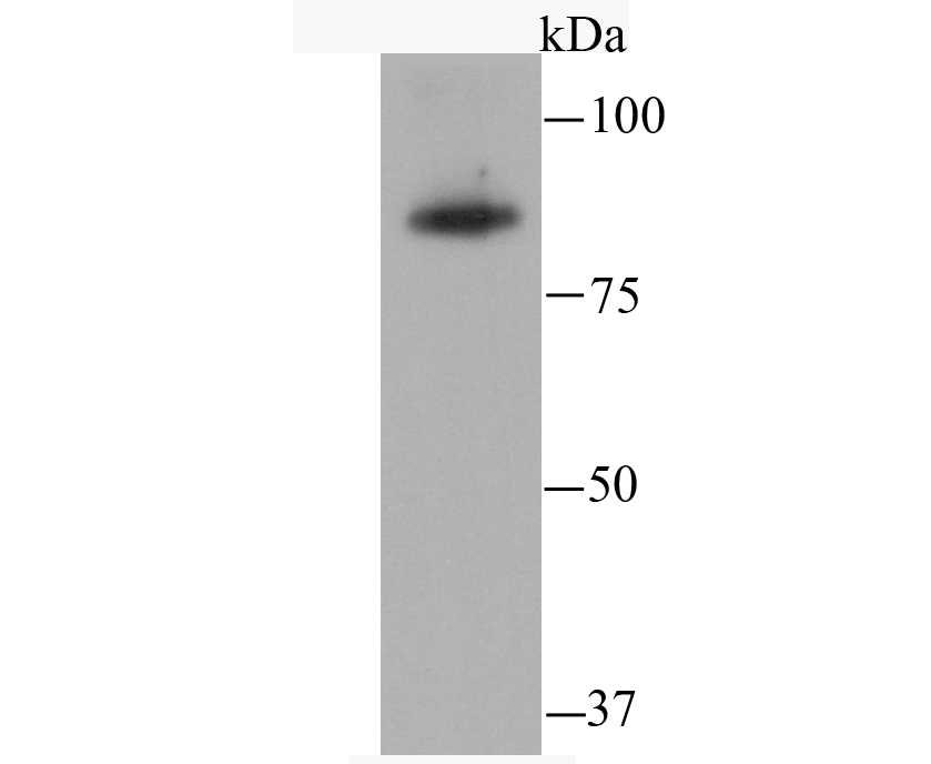
Western blot analysis of SATB2 on THP-1 cell using anti-SATB2 antibody at 1/500 dilution.
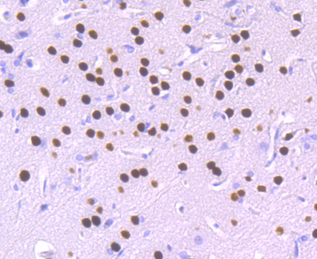
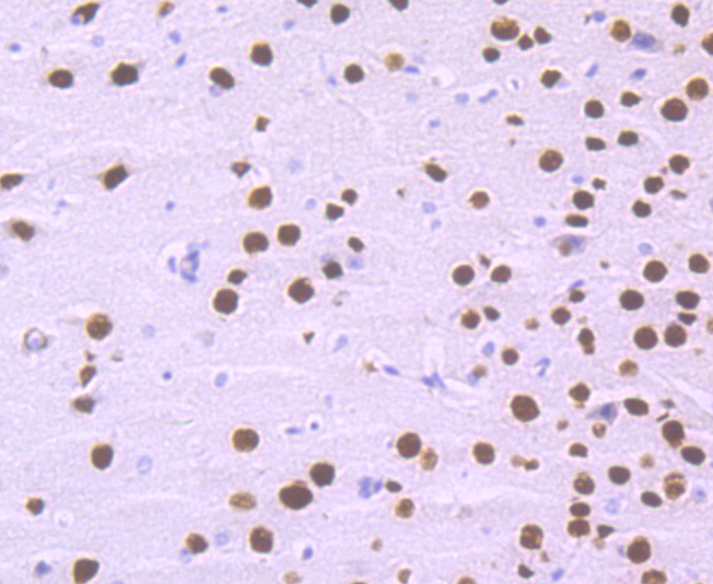
Immunohistochemical analysis of paraffin-embedded rat brain tissue using anti-SATB2 antibody. Counter stained with hematoxylin.
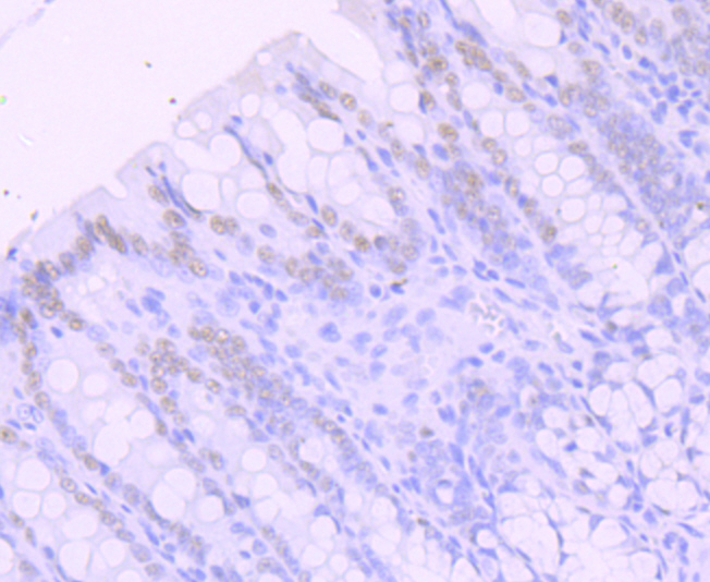
Immunohistochemical analysis of paraffin-embedded rat large intestine tissue using anti-SATB2 antibody. Counter stained with hematoxylin.
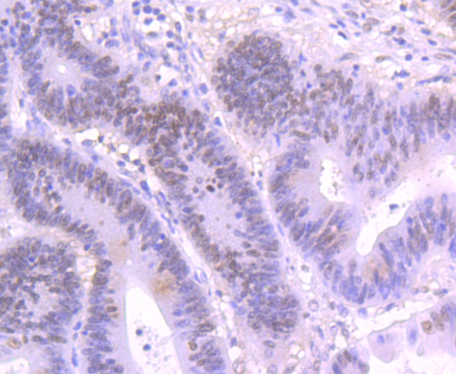
Immunohistochemical analysis of paraffin-embedded human colon cancer tissue using anti-SATB2 antibody. Counter stained with hematoxylin.
Immunohistochemical analysis of paraffin-embedded mouse brain tissue using anti-SATB2 antibody. Counter stained with hematoxylin.
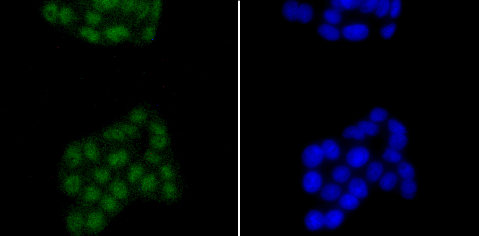
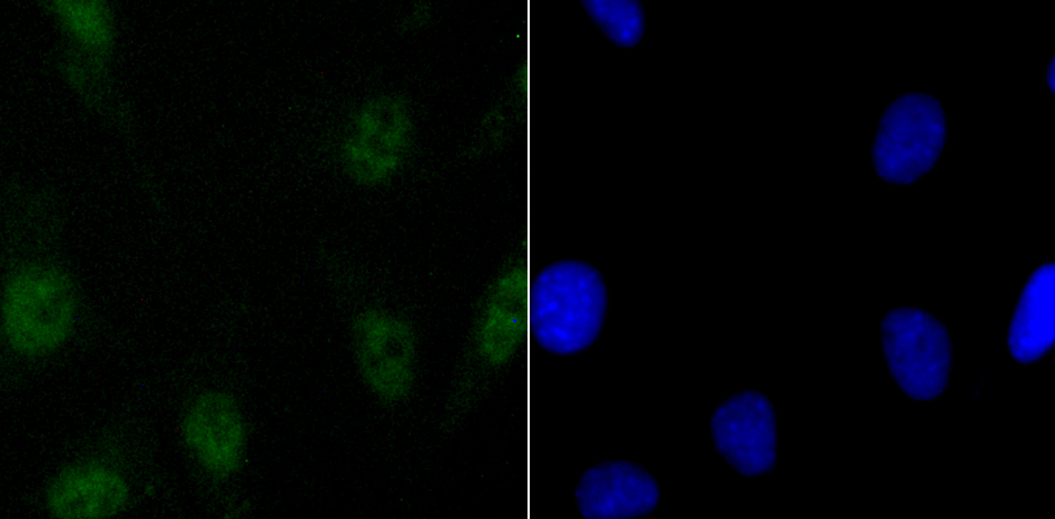
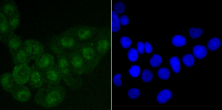
ICC staining SATB2 in PC-12 cells (green). The nuclear counter stain is DAPI (blue). Cells were fixed in paraformaldehyde, permeabilised with 0.25% Triton X100/PBS.
ICC staining SATB2 in SH-SY5Y cells (green). The nuclear counter stain is DAPI (blue). Cells were fixed in paraformaldehyde, permeabilised with 0.25% Triton X100/PBS.
ICC staining SATB2 in LOVO cells (green). The nuclear counter stain is DAPI (blue). Cells were fixed in paraformaldehyde, permeabilised with 0.25% Triton X100/PBS.
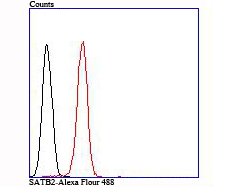
Flow cytometric analysis of SH-SY5Y cells with SATB2 antibody at 1/100 dilution (red) compared with an unlabelled control (cells without incubation with primary antibody; black).
SATB2 (Special AT-rich sequence-binding protein 2) is a nuclear matrix protein that influences craniofacial formation mechanisms, such as jaw and palate development, and is part of a transcriptional network regulating skeletal development and osteoblast differentiation. Highly expressed in adult and fetal brain, SATB2 contains two CUT DNA-binding domains and one homeobox domain and is closely related to SATB1, a transcriptional repressor. SATB2 is thought to bind to matrix-attachment regions (MARs) and regulate MAR-dependent transcription of various genes, including HoxA2 and ATF4 (CREB-2), involved in skeletal development. Functioning as both a transcriptional activator and repressor, SATB2 can also act as a protein scaffold that can enhance the activity of other DNA-binding proteins. Defects in the gene encoding SATB2 are the cause of cleft palate manifested in conjunction with severe mental retardation.
If you have published an article using product 49685, please notify us so that we can cite your literature.


 Yes
Yes


