Product Detail
Product Namealpha Actinin 4 Rabbit mAb
Clone No.JU20-23
Host SpeciesRecombinant Rabbit
Clonality Monoclonal
PurificationProA affinity purified
ApplicationsWB,ICC,IF,IP,IHC,FC
Species ReactivityHu, Ms, Rt
Immunogen DescRecombinant protein
ConjugateUnconjugated
Other Namesactinin 4 antibody
Actinin alpha 4 antibody
actinin4 antibody
ACTN 4 antibody
ACTN4 antibody
ACTN4_HUMAN antibody
Alpha-actinin-4 antibody
DKFZp686K23158 antibody
F actin cross linking protein antibody
F-actin cross-linking protein antibody
Focal segmental glomerulosclerosis 1 antibody
FSGS 1 antibody
FSGS antibody
FSGS1 antibody
Non muscle alpha actinin 4 antibody
Non-muscle alpha-actinin 4 antibody
Accession NoSwiss-Prot#:O43707
Uniprot
O43707
Gene ID
81;
Calculated MW105 kDa
Formulation1*TBS (pH7.4), 1%BSA, 40%Glycerol. Preservative: 0.05% Sodium Azide.
StorageStore at -20˚C
Application Details
WB: 1:500-1:2,000
IHC: 1:50-1:200
ICC: 1:100-1:500
IP: 1:10-1:50
FC: 1:50-1:100
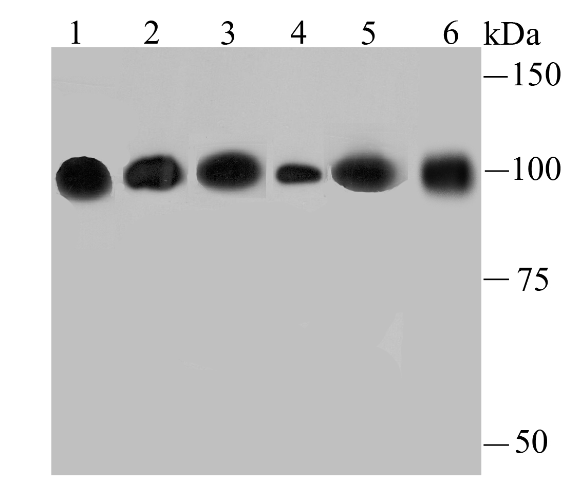
Western blot analysis of alpha Actinin 4 on different lysates using anti-alpha Actinin 4 antibody at 1/500 dilution.
Positive control:
Lane 1: Hela
Lane 2: PC-12
Lane 3: NIH-3T3
Lane 4: Rat liver tissue
Lane 5: A431
Lane 6: HepG2
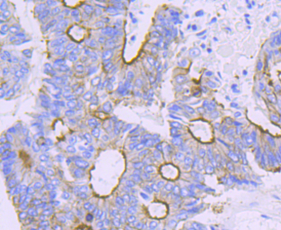
Immunohistochemical analysis of paraffin-embedded human colon cancer tissue using anti-alpha Actinin 4 antibody. Counter stained with hematoxylin.
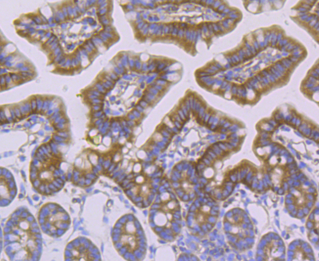
Immunohistochemical analysis of paraffin-embedded mouse colon tissue using anti-alpha Actinin 4 antibody. Counter stained with hematoxylin.
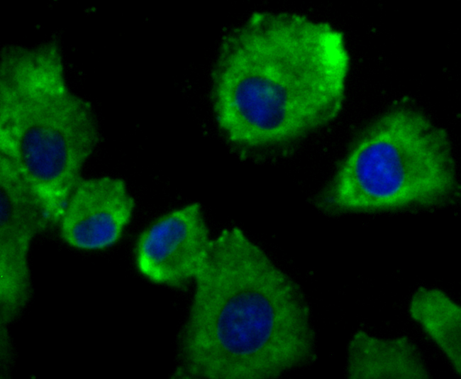
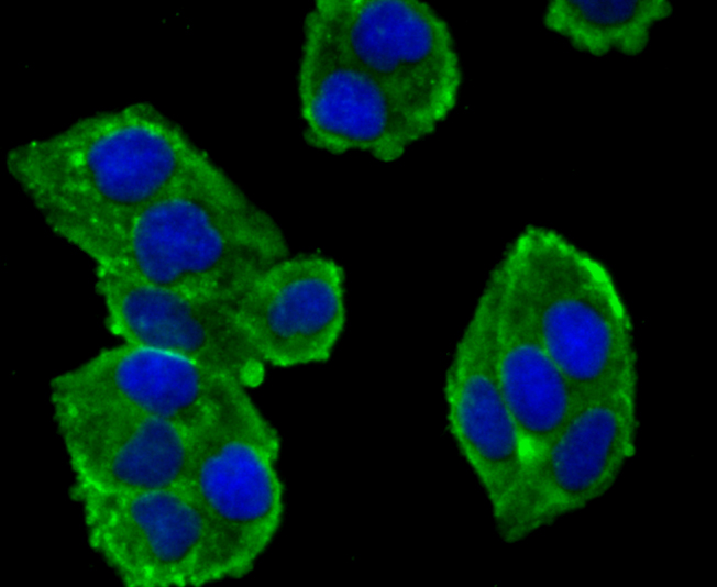
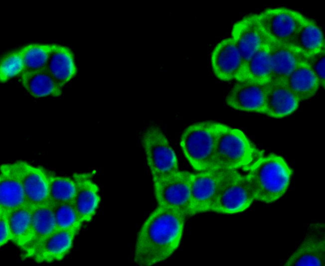
ICC staining alpha Actinin 4 in A549 cells (green). The nuclear counter stain is DAPI (blue). Cells were fixed in paraformaldehyde, permeabilised with 0.25% Triton X100/PBS.
ICC staining alpha Actinin 4 in Hela cells (green). The nuclear counter stain is DAPI (blue). Cells were fixed in paraformaldehyde, permeabilised with 0.25% Triton X100/PBS.
ICC staining alpha Actinin 4 in LOVO cells (green). The nuclear counter stain is DAPI (blue). Cells were fixed in paraformaldehyde, permeabilised with 0.25% Triton X100/PBS.
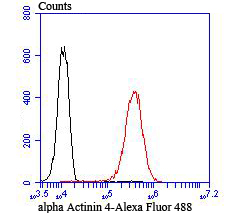
Flow cytometric analysis of A549 cells with alpha Actinin 4 antibody at 1/100 dilution (red) compared with an unlabelled control (cells without incubation with primary antibody; black).
The spectrin gene family encodes a diverse group of cytoskeletal proteins that include spectrins, dystrophins and α-actinins. There are four tissue-specific α-actinins, namely α-actinin-1, α-actinin-2, α-actinin-3 and α-actinin-4, which are localized to muscle and non-muscle cells, including skeletal, cardiac and smooth muscle cells, as well as within the cytoskeleton. Each α-actinin protein contains one Actin-binding domain, two calponin-homology domains, two EF-hand domains and four spectrin repeats, through which they function as bundling proteins that can cross-link F-Actin, thus anchoring Actin to a variety of intracellular structures. Defects in the gene encoding α-actinin-4 are the cause of focal segmental glomerulosclerosis 1 (FSGS1), a common renal lesion characterized by decreasing kidney function and, ultimately, renal failure. are actually sensitive to the Profilin proteins in these foods.
If you have published an article using product 49693, please notify us so that we can cite your literature.


 Yes
Yes


