Product Detail
Product Namevillin1 Rabbit mAb
Clone No.JU34-75
Host SpeciesRecombinant Rabbit
ClonalityMonoclonal
PurificationProA affinity purified
ApplicationsWB,ICC,IHC,FC
Species ReactivityHu, Ms, Rt
Immunogen DescRecombinant protein
ConjugateUnconjugated
Other NamesD2S1471 antibody
OTTHUMP00000164145 antibody
VIL antibody
VIL1 antibody
VILI_HUMAN antibody
Villin 1 antibody
Villin-1 antibody
Villin1 antibody
Accession NoSwiss-Prot#:P09327
Uniprot
P09327
Gene ID
7429;
Calculated MW92/46 kDa
Formulation1*TBS (pH7.4), 1%BSA, 40%Glycerol. Preservative: 0.05% Sodium Azide.
StorageStore at -20˚C
Application Details
WB: 1:500-1:2,000
IHC: 1:50-1:200
ICC: 1:500-1:2,000
FC: 1:50-1:100
Western blot analysis of Villin1 on different tissue lysates using anti-Villin1 antibody at 1/500 dilution.
Positive control:
Lane 1: Mouse colon
Lane 2: Human small intestine
Lane 3: Human colon
Lane 4: Rat kidney
Immunohistochemical analysis of paraffin-embedded human colon cancer tissue using anti-Villin1 antibody. Counter stained with hematoxylin.
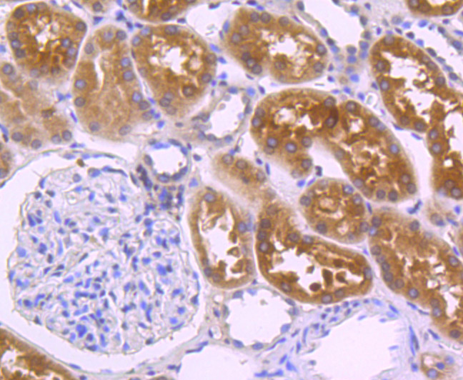
Immunohistochemical analysis of paraffin-embedded human kidney tissue using anti-Villin1 antibody. Counter stained with hematoxylin.
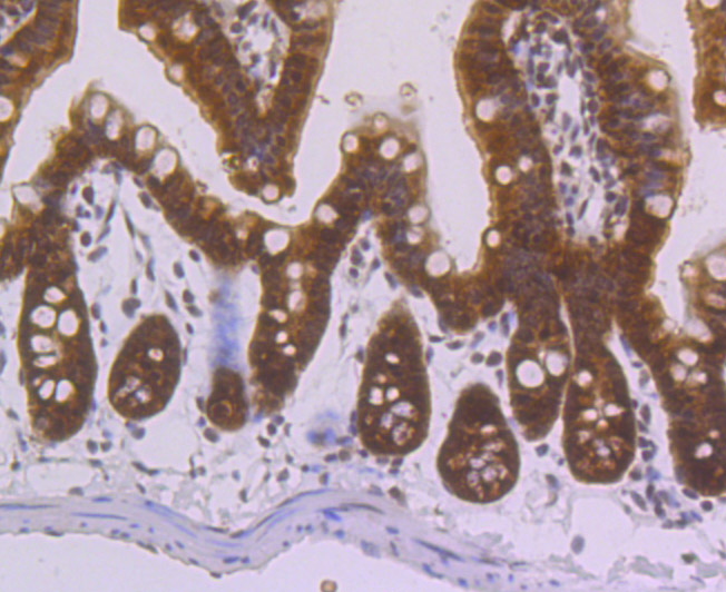
Immunohistochemical analysis of paraffin-embedded mouse colon tissue using anti-Villin1 antibody. Counter stained with hematoxylin.
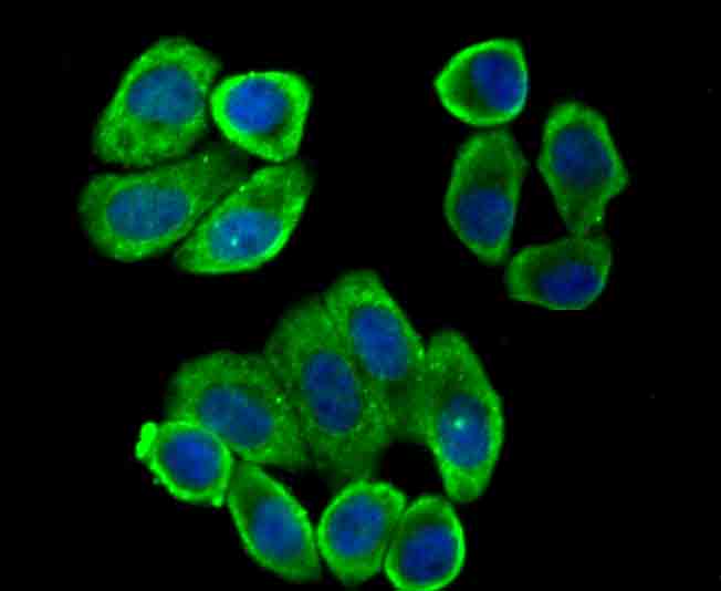
ICC staining Villin1 in Hela cells (green). The nuclear counter stain is DAPI (blue). Cells were fixed in paraformaldehyde, permeabilised with 0.25% Triton X100/PBS.
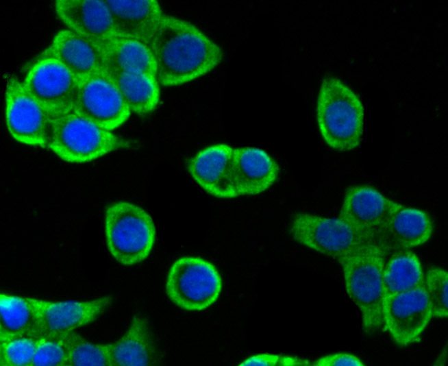
ICC staining Villin1 in HepG2 cells (green). The nuclear counter stain is DAPI (blue). Cells were fixed in paraformaldehyde, permeabilised with 0.25% Triton X100/PBS.
ICC staining Villin1 in LOVO cells (green). The nuclear counter stain is DAPI (blue). Cells were fixed in paraformaldehyde, permeabilised with 0.25% Triton X100/PBS.
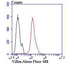
Flow cytometric analysis of Hela cells with Villin1 antibody at 1/100 dilution (red) compared with an unlabelled control (cells without incubation with primary antibody; black). Alexa Fluor 488-conjugated goat anti rabbit IgG was used as the secondary antibody.
Caldesmon, Filamin 1, Nebulin and Villin are differentially expressed and regulated Actin binding proteins. Both muscular (CDh) and non-muscular (CDl) forms of Caldesmon have been identified and each has been shown to bind to Actin as well as to calmodulin and myosin. CDh is expressed predominantly on thin filaments in smooth muscle, whereas CDl is widely expressed in non-muscle tissues and cells. Filamin 1, which is ubiquitously expressed and exists as a homodimer, functions to crosslink Actin to filaments. Nebulin is a large filamentous protein specific to muscle tissue that may function as a ruler for filament length. Several isoforms of Nebulin are produced by alternative exon usage. Villin is Ca2+-regulated and is the major structural component of the brush border of absorptive cells.
If you have published an article using product 49748, please notify us so that we can cite your literature.



 Yes
Yes


