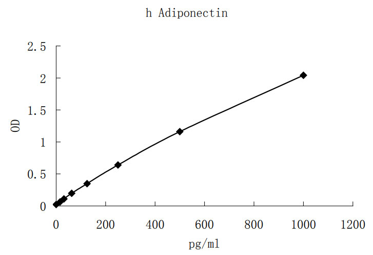
Adiponectin, alternately named Adipocyte complement-related protein of 30 kDa (Acrp30), adipoQ, adipose most abundant gene transcript 1 (apM1), and gelatin-binding protein of 28 kDa (GBP28), is an adipocyte-specific, secreted protein with potential roles in glucose and lipid homeostasis. Circulating Adiponectin levels are high, accounting for approximately 0.01% of total plasma protein (1-4). Adiponectin contains a modular structure that includes an N-terminal collagen-like domain followed by a C-terminal globular domain with significant sequence and structural resemblance to the complement factor C1q (1, 5, 6). Although they share little sequence identity, similar three-dimensional structure and certain conserved amino acid residues suggest an evolutionary link between the C1q-like domain of Adiponectin and members of the TNF superfamily (7). Adiponectin assembles into different complexes including trimers (low molecular weight), hexamers (middle molecular weight), and higher order oligomeric structures (high molecular weight) that may affect biological activity (1, 7, 8). Adiponectin is induced during adipocyte differentiation and its secretion is stimulated by insulin (1, 9). Two receptors for Adiponectin, termed AdipoR1 and AdipoR2, have been cloned (10). Although functionally distinct from G-protein-coupled receptors, the genes encode predicted proteins containing 7 transmembrane domains. AdipoR1 is highly expressed in skeletal muscle, while AdipoR2 is primarily found in hepatic tissues.
Injection of Adiponectin into non-obese diabetic mice leads to an insulin-independent decrease in glucose levels (11). This is likely due to insulin-sensitizing effects involving Adiponectin regulation of triglyceride metabolism (11). A truncated form of Adiponectin (gAdiponectin) containing only the C-terminal globular domain has been identified in the blood, and recombinant gAdiponectin has been shown to regulate weight reduction as well as free fatty acid oxidation in mouse muscle and liver (2, 12). The full-length recombinant Adiponectin protein is apparently less potent at mediating these effects (2, 12). The mechanism underlying the role of Adiponectin in lipid oxidation may involve the regulation of expression or activity of proteins associated with triglyceride metabolism including CD36, acyl CoA oxidase, AMPK, and PPARγ (12-14).
Although Adiponectin-regulation of glucose and lipid metabolism in humans is less clear, similar mechanisms may also be in place (15). A negative correlation between obesity and circulating Adiponectin has been well established (6, 16, 17), and Adiponectin levels increase concomitantly with weight loss (18). Decreased Adiponectin levels are associated with insulin resistance and hyperinsulinemia, and patients with type-2 diabetes are reported to exhibit decreased circulating Adiponectin (19, 20). Thiazolidinediones, a class of insulin-sensitizing, anti-diabetic drugs, elevate Adiponectin in insulin-resistant patients (21). In addition, high Adiponectin levels are associated with a reduced risk of type-2 diabetes (22). Using magnetic resonance spectroscopy it has been demonstrated that intracellular lipid content in human muscle negatively correlates with Adiponectin levels, potentially due to Adiponectin-induced fatty acid oxidation (15).
Adiponectin may also play anti-atherogenic and anti-inflammatory roles. Adiponectin plasma levels are decreased in patients with coronary artery disease (20). Furthermore, neointimal thickening of damaged arteries is exacerbated in Adiponectin-deficient mice and is inhibited by exogenous Adiponectin (23). Adiponectin inhibits endothelial cell expression of adhesion molecules in vitro, suppressing the attachment of monocytes (24). In addition, Adiponectin negatively regulates myelomonocytic progenitor cell growth and TNF-α production in macrophages (25, 26).
Scherer, P.E. et al. (1995) J. Biol. Chem. 270:26746.
Fruebis, J. et al. (2001) Proc. Natl. Acad. Sci. USA 98:2005.
Berg, A.H. et al. (2002) Trends Endocrinol. Metab. 13:84.
Arita, Y. et al. (1999) Biochem. Biophys. Res. Commun. 257:79.
Maeda, K. et al. (1996) Biochem. Biophys. Res. Commun. 221:286.
Kishore, U. and K.B. Reid (2000) Immunopharmacology 49:159.
Pajvani, U.B. et al. (2003) J. Biol. Chem. 278:9073.
Tsao, T.S. et al. (2003) J. Biol. Chem. 278:50810.
Hu, E. et al. (1996) J. Biol. Chem. 271:10697.
Yamauchi, T. et al. (2003) Nature 423:762.
Berg, A.H. et al. (2001) Nat. Med. 7:947.
Yamauchi, T. et al. (2001) Nat. Med. 7:941.
Tomas, E. et al. (2002) Proc. Natl. Acad. Sci. USA 99:16309.
Yamauchi, T. et al. (2002) Nat. Med. 8:1288.
Thamer, C. et al. (2002) Horm. Metab. Res. 34:646.
Stefan, N. et al. (2002) J. Clin. Endocrinol. Metab. 87:4652.
Matsubara, M. et al. (2002) Eur. J. Endocrinol. 147:173.
Faraj, M. et al. (2003) J. Clin. Endorinol. Metab. 88:1594.
Weyer, C. et al. (2001) J. Clin. Endocrinol. Metab. 86:1930.
Hotta, K. et al. (2000) Arterioscler. Thromb. Vasc. Biol. 20:1595.
Maeda, N. et al. (2001) Diabetes 50:2094.
Spranger, J. et al. (2003) Lancet 361:1060.
Matsuda, M. et al. (2002) J. Biol. Chem. 277:37487.
Ouchi, N. et al. (1999) Circulation 100:2473.
Yokota, T. et al. (2000) Blood 96:1723.
Ouchi, N. et al. (2001) Circulation 103:1057.

