Home
Antibodies
Proteins
Assay Kits
ELISA Kits
KD/KO Validation
About Us
Enzymes
Custom Service

Bax Antibody #24250

Review:
Datasheet PDF
add to favorites
Catalog No. Package Size Price Quantity
24250 100ul $529.00
Availability:Yes
Total:
ADD TO CART
 
Datasheet Images Protocol
Description
  • Product NameBax Antibody
  • Host SpeciesRabbit
  • ClonalityPolyclonal
  • PurificationIon exchange chromatography purified
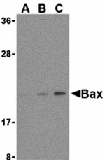
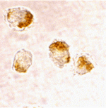
  • ApplicationsELISA WB ICC IHC
  • Species ReactivityHu Ms Rt
  • Immunogen TypePeptide
  • Immunogen DescriptionRaised against a peptide corresponding to 16 amino acids near the amino-terminus of human Bax.
  • Target NameBax
  • Accession No. Swiss-Prot:Q07812
    Gene ID:581
  • UniprotQ07812
  • Gene ID581;
  • Concentration 1mg/ml
  • Formulation Supplied in PBS containing 0.02% sodium azide.
  • Storage Can be stored at -20˚C, stable for one year. As with all antibodies care should be taken to avoid repeated freeze thaw cycles. Antibodies should not be exposed to prolonged high temperatures.
Background

Published Papers

BAD overexpression inhibits cell growth and induces apoptosis via mitochondrial-dependent pathway in non-small cell lung cancer.In Cancer Cell Int on 2013 Jun 1 by Jiang L, Luo M, et al..PMID:23725574, , (2013),
PMID: 23725574
Please let us know if you have published research using #24250 so that we can cite your reference.

Customer Reviews and Rankings

Anonymous user 05-30-2014 11:57:32

hello
dear manager: i have bout some of your product such as bax(24250) and bcl2 antibody, I want to detect Bax and bcl2 in rat brain tissue by ELISA. please send me a protocol for elisa method.
best regard
Yasin

AdministratorDear Yasin,
The following is the protocol for Elisa method.

Antigen Preparation
1. The working peptide antigen concentration is 10μg/mL: Dissolve 1mg peptide in 200μL
DI H2O (5mg/mL). Add 2μL peptide to 1mL of 50mM carbonate buffer.
2. The working protein antigen concentration is 40μg/mL: Concentrate protein to 4mg/mL
and dialyze against 50mM carbonate buffer overnight. Then dilute the protein to
40ug/mL with carbonate buffer.
Coating
3. Add 100μL of antigen at the recommended dilutions above to each well on the plate.
Incubate at room temperature for 24 hours in a humid chamber.
a. Humid chamber: Place wet paper towels in the bottom of a bin and stack the
plates on top of the towels. Cover with a lid.
Blocking
4. Remove coating solution and rinse twice (2) with DI H2O
5. Flip dry on a stack of paper towels – slap to remove all traces of liquid from the wells.
6. Add 280μL blocking buffer to each well. Incubate in a humid chamber at room
temperature for two (2) hours.
a. More blocking buffer is better, about 200‐300μL per well, but make sure not to
overfill each well.
Serum Sample
7. Dilute serum (pre‐bleed and bleed in separate Eppendorf tubes) with blocking buffer to
1:1000 (1μL serum to 1mL blocking buffer).
8. Add 125μL of 1:1000 diluted pre‐immune serum to row A of the plate.
9. Using a twelve‐channel pipettor, take 25μL from row A and add to row B, mixing with
the pipettor. Repeat with rows B and C, removing 25μL from row B and mixing it with
row C. Remove 25μL from row C and discard.
10. Add 125μL of 1:1000 diluted serum to row D of the plate.
11. Using a twelve‐channel pipettor make a series of 5 dilutions for rows E through H as
described above for the pre‐bleed, discarding 25μL after row H.
12. Incubate for one hour at room temperature.
Secondary Antibody
13. Dispose of the serum samples and wash the wells with the wash buffer using a wash
bottle or an ELISA plate washer one (1) time. Follow with one wash using DI H2O. Drain
the remaining liquid on a stack of paper towels.
14. Dilute the HRP conjugated secondary to the dilution recommended below with the
blocking buffer.
a. Goat anti‐rabbit IgG: 1:20000
b. anti‐chicken IgY: 1:5000
c. anti‐goat IgG: 1:10000
d. anti‐mouse IgG: 1:5000
e. Note: Dilutions are only recommendations. Optimal dilutions will vary for each
secondary and should be determined for each new lot.
15. Incubate at room temperature for one (1) hour.
Development
16. Dispose of the secondary and wash the wells with the wash buffer one (1) time. Follow
with one wash using DI H2O. Drain the remaining liquid on a stack of paper towels.
17. Add TMB per manufacturer instructions.
a. Positive wells will change to a blue color depending on signal intensity.
18. Add 90μL of 1M HCl to each well (or recommended media) to stop the color
development and read immediately on a microtiter plate reader at A450.
a. The blue will change to yellow when the reaction is stopped.
19. Read multiple plates in the order the color development was stopped.
It is not recommended to have more than two plates developing at the same time. Have two
timers so that the reaction is stopped after 5 minutes.

Please let us know if you have any more questions.

Best regard,
Jason

Total 1 results, divided into1 pages. First Prev Next Last

My Review

Username:Anonymous user
E-mail:
Ratings:
Content:
Verification code:captcha

Application

WBWestern Blotting

IHCImmunohistochemistry

IFImmunofluorescence

ICCImmunocytochemistry

FCFlow Cytometry

IPImmunoprecipitation

EELISA

DBDot Blotting

ChIPChromatin Immunoprecipitation

GICAGold Immunochromatography Assay

NCNegative Control

Species Reactivity

HuHuman

MsMouse 

RtRat 

Dm Drosophila melanogaster

C Caenorhabditis elegans

MkMonkey

RbRabbit

B Bovine 

D Dog

PPig

HmHamster

ChHm Chinese Hamster 

ChkChicken  

ShpSheep